
The Pentacam HR (Oculus Optikgeräte; Figure 1) is a high-resolution version of the familiar Pentacam, with optimized optics to improve its Scheimpflug anterior segment imaging capabilities. Not an optical biometer, this device nevertheless can provide vital information for planning cataract and refractive surgeries, according to the company.1 In less than 2 seconds, the device gathers diagnostic data on the entire anterior segment. The rotating scan, unlike conventional corneal topographers, measures true elevation rather than just corneal curvature; it also gathers more data points from the central cornea than conventional topography.
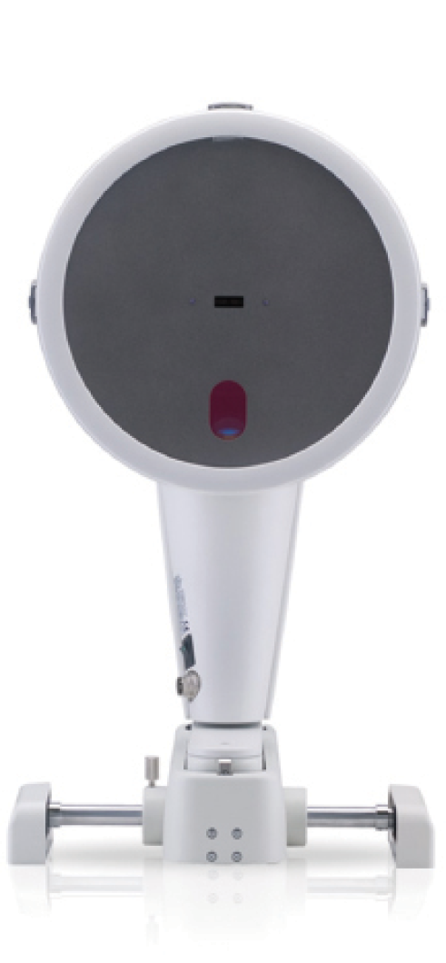
Figure 1. The Pentacam HR is a high-resolution version of the familiar Pentacam.
The device has multiple functions:
Function No. 1. Corneal and crystalline lens density are determined by light scattering and automatically quantified by the Pentacam HR's software.
Function No. 2. Color-coded corneal densitometry maps allow customized and standardized evaluation of the full depth and breadth of the cornea.
Function No. 3. Analysis of corneal optical density allows detection of corneal pathology such as Fuchs dystrophy and corneal scars.
Function No. 4. Measurements of both anterior and posterior corneal surfaces enable calculation of the true refractive power of the cornea and facilitate planning for toric IOL power and orientation. This function also can detect early ectatic changes.
The optimized optics of the Pentacam HR provide bright, high-quality images, with five times greater resolution than standard Pentacam models. This means that corneal irregularities are made visible, as are signs of trauma or refractive interventions such as corneal implants or incisions. Additionally, a cornea fine-scan mode can be used to view intracorneal pathologies. The device can also show detailed images of phakic and pseudophakic IOLs in situ.
In addition to the basic Pentacam software, which includes reports for glaucoma and refractive surgery screenings, the available contact lens fitting software incorporates Fourier analysis. Other features include the iris camera, providing automatic horizontal white-to-white measurement, and enhanced reports including topography maps of the anterior and posterior corneal surfaces; elevation maps of these surfaces; absolute and relative pachymetry maps; 3-D anterior chamber analysis, providing chamber volume and depth and the anterior chamber angle; anterior segment topography; and topometric keratoconus detection and classification. On comparative displays, up to four exams can be loaded and analyzed to show progression over time, and two exams can be shown side by side—convenient for comparing differences in left and right eyes.
The device can link to several online IOL power calculation programs, including the i-ASSORT (ASSORT), BESSt II (EB-EYE), Holladay IOL Consultant & Surgical Outcomes Assessment (Holladay Consulting), and PhacoOptics (IOL Innovations). A floating license allows the Pentacam software to be used simultaneously on multiple workstations linked on a practice's local network. Optional software packages can be included on the floating license. n
1. The Pentacam. Oculus website. http://www.pentacam.com/sites/hr.php. Accessed June 22, 2015.