We noticed you’re blocking ads

Thanks for visiting CRSTG | Europe Edition. Our advertisers are important supporters of this site, and content cannot be accessed if ad-blocking software is activated.

In order to avoid adverse performance issues with this site, please white list https://crstodayeurope.com in your ad blocker then refresh this page.

Need help? Click here for instructions.

Cover Focus | Nov/Dec 2015

SMILE: A Brief Review

An example of one intrastromal procedure that can be added to the refractive surgeon's arsenal.

Small incision lenticule extraction (SMILE), performed using a femtosecond laser (VisuMax; Carl Zeiss Meditec) that is capable of carving out a lenticule within the cornea, is one form of intrastromal refractive surgery. The laser energy of the VisuMax creates plasma-induced photodisruption, resulting in the formation of cleavage planes within the corneal stroma at a predetermined depth with a high degree of precision.1 The lenticule can then be extracted from within the corneal stroma by creating and lifting a hinged flap in a procedure called femtosecond lenticule extraction (FLEX) or via the more popular SMILE procedure, in which the lenticule is extracted through a small incision.

Figure 1. In Circle, a lamellar ring is created at the same depth as the existing SMILE incision, allowing the conversion of the SMILE cap into a hinged flap. The flap is then raised, and excimer laser ablation is performed.

SMILE has become an alternative to LASIK for myopic correction for many surgeons in Europe and Asia (see The Three Phases of SMILE on the previous page).2,3 It is awaiting FDA approval in the United States pending the results of ongoing clinical trials. The procedure is currently available as a treatment modality for myopic correction of up to -10.00 D spherical equivalent, with a maximum astigmatic error of up to 5.00 D. The selection criteria are similar to those for LASIK. The procedure is currently not capable of hyperopic correction, although research is ongoing for this application.

MANAGING COMPLICATIONS

There are two types of complications that can be encountered intraoperatively, and each can potentially be managed successfully.

Suction loss. This occurs when the contact glass becomes detached from the cornea during the procedure.4 The general challenge in this situation is redocking of the contact glass interface to the eye while still retaining centration. In our experience, repeating the treatment immediately is convenient and does not seem to affect the results of the procedure.

The Three Phases of SMILE

Phase 1: Initial docking with precise centration. During this phase, proper head position is achieved by tilting the patient's head medially to avoid nasal contact with the cone of the contact glass interface. Precise centration should be verified before the initiation of suction.

Phase 2: Femtosecond laser delivery. Femtosecond laser pulses are fired in a spiral pattern with a pulse energy of 120 to 170 nJ and a repetition rate of 500 kHz.

Phase 3: Tissue dissection and lenticule removal. The tissue disruption planes created by the laser include the posterior lenticule surface, vertical edge cut, anterior lenticule surface, and corneal sidecut (Figures 1 and 2).

Figure 1. Cross-section of the cleavage planes created during SMILE: (1) posterior lenticule surface, (2) vertical edge cut, (3) anterior lenticule surface, and (4) corneal sidecut.

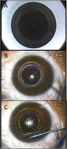
Figure 2. Surgical steps of the SMILE procedure, including laser-assisted and manual steps. Laser-assisted: posterior tissue disruption plane, or lenticule cut (A); anterior tissue disruption plane, or flap cut (B); superior flap and sidecut incision (C). Manual steps: delineation of planes (D), dissection of planes (E), and lenticule removal (F).

Incorrect dissection. Here, the posterior disruption plane is separated first, resulting in the lenticule being stuck to the undersurface of the cap. In our experience, when this occurs it is still possible, with some surgical dexterity, to separate the lenticule from the overlying cornea. In case this is not possible, the VisuMax allows the creation of a sidecut incision only, and it is best at this point to convert the case into a FLEX procedure by repeating a 270º to 300° sidecut incision.

SMILE OVER LASIK

Several authors have demonstrated superiority of SMILE over femtosecond LASIK in a number of measures. SMILE has been reported to result in lower degrees of induced higher-order aberrations and less reduction in corneal sensitivity than LASIK.5,6

Also, SMILE affects the biomechanical properties of the cornea less than LASIK.7,8 SMILE is a flapless technique, and its effect is predicated on the new concept of tissue subtraction, which is different from tissue ablation achieved with the excimer laser in LASIK.7,8

enhancement technique

No matter how accurate the results of SMILE, no doubt some patients will need enhancement. A recent adaptation of the VisuMax software enables enhancements of SMILE with a procedure known as Circle (Figure 1). With Circle, a previously created cap can be remodeled into a larger diameter hinged flap, and this can be followed by excimer laser ablation to address any residual refractive error. n

1. Sekundo W, Kunert K, Russmann C, et al. First efficacy and safety study of femtosecond lenticule extraction for the correction of myopia: 6 month results. J Cataract Refract Surg. 2008;34:1513-1520.

2. Sekundo W, Kunert K, Blum M. Small incision corneal refractive surgery using the small incision lenticule extraction (SMILE) procedure for the correction of myopia and myopic astigmatism: results of a 6 month prospective study. Br J Ophthalmol. 2011;95:335-339.

3. Shah R, Shah S, Sengupta S. Results of small incision lenticule extraction: all-in-one femtosecond laser refractive surgery. J Cataract Refract Surg. 2011;37:127-137.

4. Wong CW, Chan C, Tan D, et al. Incidence and management of suction loss in refractive lenticule extraction. J Cataract Refract Surg. 2014;40:2002-2010.

5. Li X, Wang Y, Duo RV. Aberration compensation between anterior and posterior corneal surfaces after small incision lenticule extraction and femtosecond laser-assisted laser in-situ keratomileusis. Ophthalmic Physiol Opt. 2015;35(5):540-541.

6. Gyldenkerne A, Ivarsen A, Hjortdal JØ. Comparison of corneal shape changes and aberrations induced by FS-LASIK and SMILE for myopia. J Refract Surg. 2015;31(4):223-229.

7. Wu D, Wang Y, Zhang L, Wei S, Tang X. Corneal biomechanical effects: small-incision lenticule extraction versus femtosecond laser–assisted laser in situ keratomileusis. J Cataract Refract Surg. 2014;40:954-962.

8. Sinha Roy A, Dupps WJ Jr, Roberts CJ. Comparison of biomechanical effects of small-incision lenticule extraction and laser in situ keratomileusis: finite-element analysis. J Cataract Refract Surg. 2014;40:971-980.

Mahipal Sachdev, MD
• Chairman, Medical Director, and Senior Consultant Ophthalmologist, Centre For Sight, New Delhi, India
drmahipal@gmail.com
• Financial disclosure: Travel grant (Carl Zeiss Meditec)

NEXT IN THIS ISSUE