


A 30-year-old woman reported dim vision in both eyes. On examination, her BCVA was 6/9 OD with a manifest refraction of -24.00 -2.00 x 50º and 6/24 OS with a manifest refraction of -27.00 -1.50 x 140º.
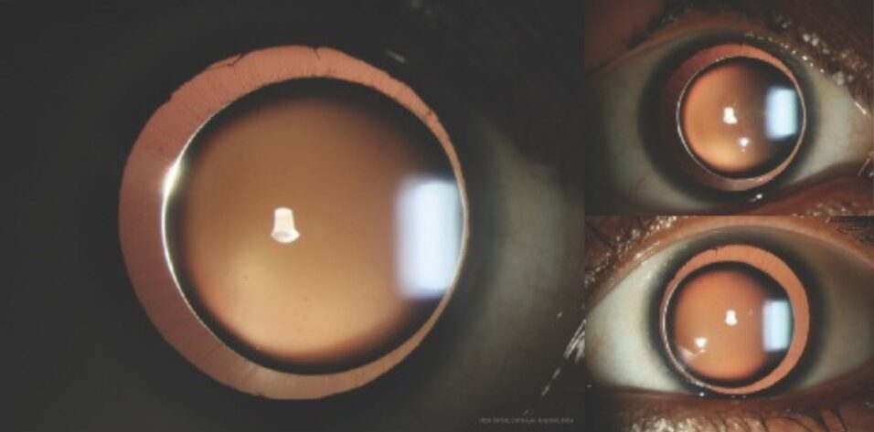
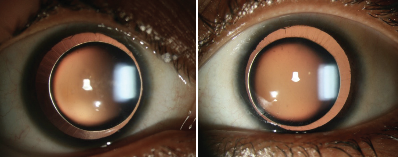
A slit-lamp examination found microspherophakia (Figures), a deep anterior chamber, and considerable phacodonesis in each eye. The IOP was 18 mm Hg OD and 20 mm Hg OS. Due to the increased anteroposterior thickness of the lens, the slit beam reflected off the posterior surface, producing a diamond ring–like effect with retroillumination that resembled a solar eclipse (main image).
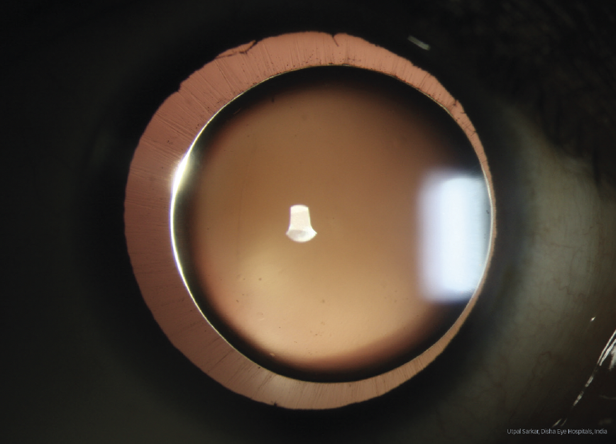
Figure. Bilateral microspherophakia with deep anterior chambers and considerable phacodonesis.
The patient was referred to the retina department. An examination was normal and identified no peripheral retinal degeneration. A diagnosis of spontaneous lens dislocation and pupillary block glaucoma was made. A vitrectomy and lensectomy under local anesthesia were recommended, but the patient declined surgery.


