


Thanks to advances in phaco technology and techniques, there are few instances when surgeons cannot crack and emulsify a cataract. Just because they can, however, doesn’t mean they should.
The repeated instillation of a dispersive OVD can help protect the cornea, but reducing the amount of ultrasound energy used during surgery may be more important. Much of the energy is used when the initial groove is created to help crack a dense nucleus. I have had success with a stop and chop technique—even in moderately dense lenses—but I prefer to use the miLoop (Carl Zeiss Meditec) when I am concerned that a posterior plate may require increased surgical time and ultrasound energy.
DECREASING PHACO ENERGY: A CASE EXAMPLE
Use of the miLoop is associated with a statistically significant reduction in ultrasound energy,1 and the device can easily break through a posterior plate that would slow and complicate manual cracking. This was particularly helpful in the case of a 66-year-old man with high myopia and a dense brown/black posttraumatic cataract from a childhood injury. The patient had previously been told by another physician that cataract surgery would not be beneficial.
After a thorough examination, I explained to the patient that his postoperative visual acuity might be limited by retinal or optic nerve pathology. He decided to proceed with cataract surgery. Attachment of the retina was confirmed with B-scan ultrasonography.
My plan was to perform small-incision extracapsular cataract extraction and to divide the nucleus into multiple segments with the miLoop, which I hoped would allow me to use a small scleral incision.
Despite dividing the lens into four small pieces with the miLoop, phacoemulsification was difficult. I had made a small extracapsular incision but did not need to extend it to the full length—only to the width of the lens loop (Figure 1)—but enlargement of the clear corneal incision and proper placement of corneal sutures can be required. If I place limbal sutures, I start to remove them 2 to 3 weeks after surgery and have thus far managed to avoid inducing significant astigmatism.
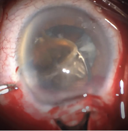
Figure 1. The lens loop is used to remove a dense cataract piece.