
Glaucoma care is more of an art than a science. The introduction of novel MIGS procedures and several new classes of IOP-lowering medications have increased management options but have done little to standardize the treatment course. Patient management remains crucial to successful intervention.
In my hands, the OMNI® Surgical System (Sight Sciences) leads to a more controllable and reproducible postoperative journey in properly selected patients. The goal is to be realistic about what can be achieved. For many patients, pressures in the mid-teens (15 to 18 mm Hg) are a success when they can stay off drops.
The OMNI Surgical System is unique in that it combines canaloplasty and trabeculotomy—two procedures with a significant evidence base—to alleviate outflow resistance in the conventional outflow pathway. It can be used to treat 360º of Schlemm’s canal, addressing three sources of outflow resistance—trabecular meshwork, Schlemm’s canal, and collector channels—in a single procedure.
GETTING STARTED
Patient selection. Like any new device or procedure, the best way to maximize the potential for good postoperative results with OMNI is to start with patients who demonstrate a mild to moderate disease state. Those who present with pigmented angles are ideal early candidates for MIGS procedures like OMNI because it is easier to identify landmarks compared to eyes with pale angles. I like to perform slit-lamp gonioscopy, separating the observation tower from the illumination tower by about 30º, to provide a crisp view of the corneal light wedge (ie, anterior-posterior corneal interface and Schwalbe's line.)
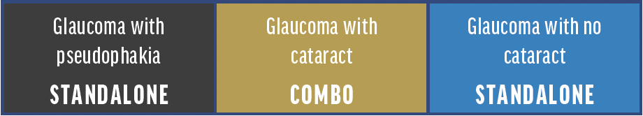
OMNI can be used in combination with cataract surgery or as a standalone procedure (see chart above). I recommend starting by performing the procedure combined with phaco or in pseudophakic patients; those who may have had SLT are still suitable. I would avoid phakic patients for the first few cases to improve access to the angle and reduce concerns about touching the crystalline lens. If the angle is tight, combining OMNI with phaco helps open the angle. A standalone procedure can be considered if access to the angle is sufficient.
Intraoperative tips. In my opinion, the learning curve with OMNI is short. One of the first lessons I learned with all MIGS devices is to position the corneal wound toward my dominant hand. This creates a more natural hand position and results in less corneal warpage, improving the gonioscopic view and minimizing OVD loss from the anterior chamber. Once the device is inserted in Schlemm’s canal, it only takes a few cases to get used to how it is advanced. The wheels should be advanced smoothly and carefully. If the catheter meets resistance or kinks momentarily, then it implies a tight area of Schlemm’s canal. Don’t force the catheter through. Retract the wheel slightly (this adds OVD expansion and lubrication) and then re-pass. It should now slide in easily without resistance.
It is important to come out of the eye with the OMNI device and turn the tip over to realign it before making a second pass to protect the lens and cornea. Cannulating forehand (ie, to the left for a right-handed surgeon) is often easier than cannulating backhand (ie, to the right for a right-handed surgeon), particularly for the first few cases.
The 11 microliter reservoir in the OMNI device can hold enough OVD for two 180º passes. Aim to tilt the OMNI handpiece upward about 30º. Once in the canal, the wire will slide horizontally (Figure). A flat position can lead to the device diving down toward the iris.
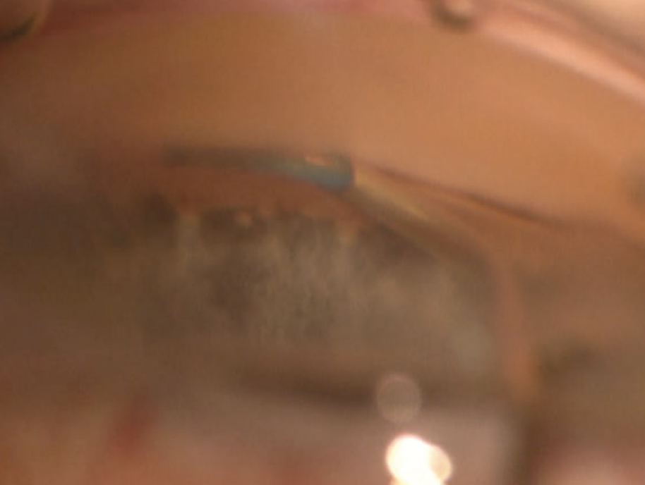
Figure. Proper placement of OMNI.
In addition to intracameral cefuroxime, I like to use intracameral dexamethasone (0.66 mg in 0.2 mL) in the anterior chamber for viscocanaloplasty-only cases. I add subconjunctival dexamethasone if I perform additional trabeculotomy.
Postoperative care. The next day after surgery, the eyes are usually quiet, helped by the intracameral steroid depot. If I perform trabeculotomy, I prefer to add pilocarpine to control inflammation and stop adhesions to the trabecular meshwork. However, because it increases blood-aqueous barrier porosity, pilocarpine is prescribed only for a short duration. Additionally, I prefer to use a little extra steroid when I treat 360º so that I don’t have any postoperative surprises. This consists of dexamethasone every 2 hours (daytime only) for the first week, tapering over 4 or 6 weeks for viscocanaloplasty and trabeculotomy, respectively. With OMNI, most patients can stop IOP-lowering medication use after surgery. I prefer to drop the prostaglandin first and reduce sequentially, one medication at a time.
CONSISTENT PRESSURES
After the OMNI procedure, in my experience, many patients can maintain consistent IOP in the mid-teens. In one study of standalone OMNI with 24-month follow-up, IOP decreased from 24.6 mm Hg on 1.9 medications to between 12.6 and 14.9 mm Hg on 0.9 medications. Most (84.6%) patients reduced their medications by 1 and still achieved a near 50% IOP reduction. Additionally, 57.7% were medication-free.1
Most of my OMNI patients have had mild to moderate open-angle glaucoma. In my experience, these patients have better early IOP than other MIGS devices.
In my practice, 6-month follow-up data is available for 37 patients at the time of this writing. The mean preoperative IOP was 24.1 mm Hg on 2.4 medications. These cases only had 360º viscocanaloplasty (no trabeculotomy); the procedure was combined with phacoemulsification in 35 of 37 eyes. At 6 months, the mean IOP improved to 14.5 mm Hg and mean medication use to 1.6. My results exceed my personal experiences of devices treating trabecular meshwork only.
CONCLUSION
Patient selection is essential for MIGS procedures, including procedures with the OMNI Surgical System. Preoperative slit-lamp gonioscopy is essential before suggesting OMNI to patients. In my experience with OMNI in mild to moderate open angle glaucoma, I can achieve reliable pressure outcomes in the mid-teens with a significant reduction or elimination of their medication burden (dependent on number of preoperative medications). Additional steroid therapy is advisable for surgeons moving to OMNI from trabecular stenting procedures.
1. Klabe K, Kaymak H. Standalone trabeculotomy and viscodilation of Schlemm’s canal and collector channels in open-angle glaucoma using the OMNI Surgical System: 24-month outcomes. Clin Ophthalmol. 2021;15:3212-3129.
Indications for use
The OMNI® Surgical System is indicated for the catheterization and transluminal viscodilation of Schlemm’s canal and the cutting of trabecular meshwork to reduce intraocular pressure in adult patients with open-angle glaucoma.
Contraindications
Do not use the OMNI in any situations where the angle is compromised or has been damaged (e.g., from trauma or surgery), since it may not be possible to visualize the angle or to properly pass the microcatheter. Do not use the OMNI in patients with angle recession; neovascular glaucoma; chronic angle closure; narrow-angle glaucoma; traumatic or malignant glaucoma; or narrow inlet canals with plateau iris.
Please refer to the full Instructions For Use, available at OMNIsurgical.com, for warnings, precautions, and adverse event information.
@ 2022 Sight Sciences, Inc. OM-2678-OUS.v1 12/22

