
Augmented Awareness: Supporting the Review Process with AI-based Anomaly Detection in Corneal OCT
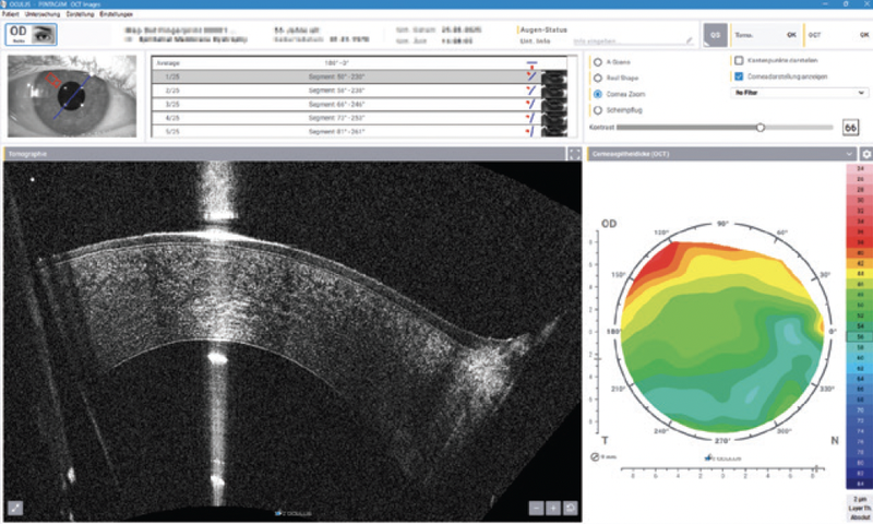
The increasing resolution of anterior segment imaging has opened new dimensions in diagnostics, but reviewing the vast amount of structural detail remains a challenge in clinical routine. To address this, a novel AI-based support tool is currently being explored for the Pentacam® Cornea OCT. Its purpose: to assist users in detecting suspicious findings within corneal OCT cross-sections without altering diagnostic responsibility.8
Supporting the Review Process
Each Pentacam® Cornea OCT exam captures 25 high-resolution B-scans across the cornea. While these scans offer exceptional insight into fine structures – including epithelial integrity, Bowman’s layer disruptions, or endothelial changes – they can be time-consuming to review thoroughly. In many clinics, this task is left to the interpreting physician, while technicians perform the scan itself.
The new AI-based support system is designed to bridge this workflow gap and reduce the likelihood of missed localized anomalies.8 After image acquisition, the system automatically analyzes all 25 B-scans and flags potential anomalies. Any image with suspected irregularities is visually marked, drawing attention to regions that may warrant closer review.
The following shows a Pentacam® Cornea OCT examination, which initially appeared normal. Thorough analysis of the 25 cross sectional images reveals epithelial abnormalities in the 18th section, showing classical changes of an epithelial basement membrane dystrophy (EBMD). This abnormality was visually marked by the algorithm, immediately indicating abnormal findings in this case. Otherwise, the EBMD might have remained undetected or only been detected after careful analysis of every single section, which takes several minutes in clinical practice.


Focused Attention, Enhanced Workflow
This feature offers several practical benefits. First, physicians can prioritize their attention on flagged sections, ensuring that subtle but important changes are not overlooked. Second, it empowers trained technicians to perform targeted follow-up scans before the patient even leaves the exam room. Averaging or Sector Scans of the area of interest provide enhanced noise-free images or imaging of anomalies with increased angular resolution, respectively, ensuring a more comprehensive and efficient imaging assessment.
In doing so, the Pentacam® Cornea OCT can help to prevent delays in care, reduce the need for repeat appointments, and strengthen the communication between technician and physician. In busy practices, where time is limited and diagnostic certainty is essential, such efficiency gains can translate into measurable improvements in patient care.
The goal is not to replace clinical judgment but to augment diagnostic awareness and streamline the transition from data acquisition to diagnostic decision-making.
A Cautious and Compliant Approach
To train the system, a large dataset of labeled OCT images was used, including both normal and pathological cases, providing the algorithm with a rich foundation for pattern recognition. However, given the evolving regulatory landscape surrounding AI in ophthalmology, this feature is currently under critical evaluation and not yet available for routine clinical use.
No diagnostic decisions are made by the system. It is designed as a supportive tool to highlight image areas for closer inspection. Clinical interpretation and decision-making remain fully under the control of the physician.
Building Confidence Through Intelligence
As OCT becomes more detailed and datasets grow larger, tools that help clinicians focus on what matters most are becoming increasingly valuable. With this AI-based assistance, the Pentacam® Cornea OCT continues to evolve – not only as a precision imaging system, but also as an intelligent support platform for diagnostic precision.9
The views and opinions expressed here may not necessarily reflect those of Bryn Mawr Communications or Cataract & Refractive Surgery Today Global.
Ready to Claim Your Credits?
You have attempts to pass this post-test. Take your time and review carefully before submitting.
Good luck!
Recommended
 See the Unseen
Redefining Corneal Tomography: Introducing the Pentacam® Cornea OCT
Renato Ambrosio, Jr., MD, PhDRenato Ambrosio, Jr., MD, PhD