


I learn from surgeons around the world every day!
Uday Devgan, MD, FACS

Ophthalmologists begin with a foundation of knowledge laid during residency and fellowship, and then we spend a lifetime building on that. The only constant in ophthalmic surgery is change, and our techniques and technologies evolve every year. If we perform surgery using only the methods we learned during residency training, we will soon find ourselves behind the times.
One of the best sources of cutting-edge surgical information is our fellow surgeons, and the best way to learn from them is to observe them in the OR. Two decades ago, I was fortunate to travel to more than 50 countries and 36 US states to learn from other surgeons, give lectures, and perform demonstrations. Over the course of many years, I learned from the best surgeons in the world, but the experience came at a cost: I was away from my own practice for 2 to 3 months per year, and I had to travel more than 1 million miles. Nevertheless, it was the best fellowship that I can imagine, and I brought newly learned surgical techniques back to my practice in Los Angeles for the benefit of my patients.
AN EVOLUTION IN LEARNING
I learned surgery from my excellent mentors at the University of California at Los Angeles Jules Stein Eye Institute and from other surgeons around the world via the Video Journal of Cataract & Refractive Surgery from Robert H. Osher, MD, (for more on this and other online surgical video resources, see Online Video Education Resources). As a resident, I searched far and wide to find VHS tapes of these videos so that I could study them carefully. Now, they are available on Eyetube.
It’s a new world. Instead of low-resolution Super VHS, videos are recorded in high-definition or even 4K, and they can be watched on a mobile phone virtually anywhere in the country. Currently, my preferred way of learning surgery is to watch surgical videos. Although they are filmed mostly from the surgeon’s view through the operating microscope, many use two camera angles and show the external view (Figure 1). This is as good as being there in person. Even if I visit a center, I watch the surgery on a video monitor in the OR because I do not have privileges to scrub into the cases.
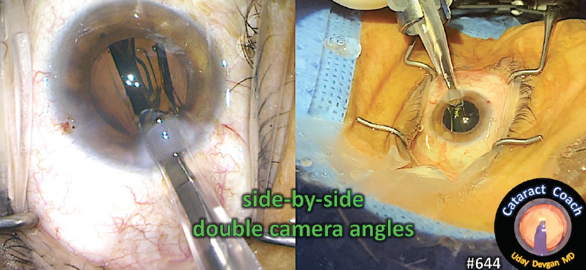
Figure 1. This is an example of the double–camera-angle videos that show the surgeon’s view through the microscope and the external view simultaneously. This creates a better learning experience and is as good as being there in person.
Three years ago, I started CataractCoach.com, which is a free surgical teaching website. It features a new video every day and is approaching video number 1,000. These are all categorized and indexed, and there is a powerful search engine that makes navigating the videos and finding a particular topic easy. In the past month alone, I have learned from dozens of ophthalmologists worldwide and in the United States on this website. Each of them has taught me an important surgical pearl that ultimately benefits my patients. The best part is that each video is just 5 minutes long.
THE POWER OF PEER EDUCATION
My surgical skill set has benefitted from watching my peers, both live and on video. Below is just a handful of surgeons who I have learned from by watching their videos (Figure 2).
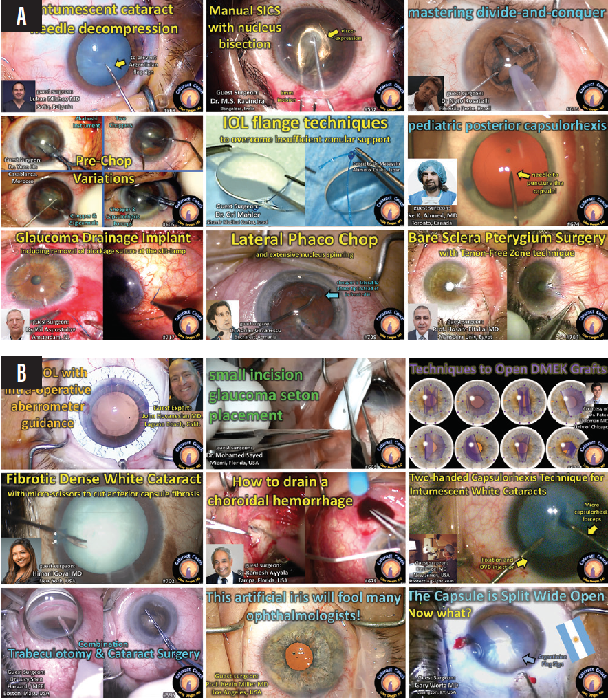
Figure 2. Colleagues from outside (A) and within the United States (B) have taught Dr. Devgan and others so much about ocular surgery from their videos on CataractCoach.com.
Figures 1 and 2 courtesy of Uday Devgan, MD, FACS
International Colleagues
- Lukan Mishev, MD, from Bulgaria, is a master surgeon who often livestreams his cases to a worldwide audience. He taught me the art of decompressing intumescent white cataracts with a needle.
- M.S. Ravindra, MD, MBBS, from India, showed me the technique of manually bisecting the nucleus while performing manual small-incision cataract surgery.
- Neto Rosaelli, MD, from Brazil, has mastered just about every cataract surgical technique, and it is a pleasure to learn from him.
- Yan Lin, MD, from Morocco, has taught me multiple prechop variations and showed me that even a blunt cannula can split a nucleus.
- Ori Maher, MD, from Israel, is the king of IOL flange techniques, and I have learned a lot from watching his cases.
- Iqbal Ike K. Ahmed, MD, FRCSC, from Canada, showed me how to succeed in pediatric cataract cases that require a posterior capsulotomy.
- Val Apostolov, MD, from the Netherlands, is a truly gifted surgeon, and I have improved my surgical technique in glaucoma cases by observing him.
- Adrian Gavanescu, MD, from Romania, showed me that the chop technique could be lateral instead of just linear.
- Hosam El-Fallal, MD, from Egypt, showed me how to improve pterygium surgery by creating a Tenon-free zone.
Colleagues in the United States
- John Hovanesian, MD, from Laguna Beach, California, is an outstanding surgeon who taught me when he was a cornea fellow and I was an ophthalmology resident at the University of California at Los Angeles more than 20 years ago. He still teaches me with every video he makes.
- Mohamed Sayed, MD, from Miami, showed me that it is possible to place a glaucoma seton device through a small incision, something I had never seen before.
- Peter Veldman, MD, from Chicago, has made me better at Descemet membrane endothelial keratoplasty by showing all of the graft configurations and how to solve the unfolding dilemma—pure genius.
- Himani Goyal, MD, from New York City, showed that anterior segment surgeons can use retinal instrumentation for tough cataract cases.
- Ramesh S. Ayyala, MD, from Tampa, Florida, taught me how to drain the blood from a choroidal hemorrhage. Although I have not yet encountered this complication, I am now prepared to deal with it.
- Ravi D. Goel, MD, from Cherry Hill, New Jersey, showed that using both hands during a challenging case will allow me to fixate the eye and inject an OVD while performing the capsulorhexis.
- Lucy Q. Shen, MD, from Boston, taught me the nuances of concurrent glaucoma and cataract surgery. I consider her surgical technique to be among the best on the planet.
- Kevin M. Miller, MD, works down the street from me in Los Angeles, but I have not visited his OR since the culmination of my residency. His video showed me a brilliant technique for artificial iris implantation.
- Gary Wörtz, MD, from Lexington, Kentucky, showed me a technique for successfully completing cataract surgery and achieving an excellent visual outcome despite being surprised by the Argentinian flag sign.
Conclusion
Traveling thousands of miles to watch another surgeon in the OR or to hear another surgeon speak at a large ophthalmology meeting seems increasingly inefficient. The future of ophthalmic surgical education is streaming videos online. We can watch these videos whenever we want and review them before our own similar surgical procedures. The most important beneficiaries of this education are our patients.
I learned a lifetime of lessons by visiting Joseph Colin, MD.
Aylin Kiliç, MD

I’ve always been interested in helping patients with keratoconus. Early in my career, there were many young, usually hopeless patients with this disease visiting my clinic. (Corneal transplantation cannot be scheduled within a short time in my country.) These patients were often contact lens intolerant and navigating life with difficulty because they were unable to see clearly. Around that time, the idea of changing the corneal shape using an intrastromal corneal ring segment (ICRS) was an exciting one. It was a creative approach: The nomogram was not clear, and there were so many aspects of the procedure to discuss and consider.

Figure 3. Drs. Kiliç and Colin at an American-European Congress of Ophthalmic Surgery meeting in Cannes, France, in their last photograph together before Dr. Colin’s death in 2013.
Courtesy of Aylin Kiliç, MD
In 2007, I was introduced to Joseph Colin, MD (Figure 3), by Jose Luís Güell, PhD. I was full of questions about how ICRSs work. Dr. Colin invited me to visit his clinic in Bordeaux, France. I gave my first international talk about femtosecond lasers at his university. It was an important topic for me because I was planning to perform ICRS surgery using a femtosecond laser. My visit to Dr. Colin’s practice was so helpful. He explained many details of the procedure and answered all of my questions. I watched him perform an ICRS operation and peppered him with questions about tunnel size, ring size, astigmatism management, tunnel depth, indications, and the importance of the ICRS. He graciously answered them all.
At that time, PMMA was being used for ICRSs. Dr. Colin told me, “The cornea never accepts a foreign body. There will be rejection.” Afterward, I focused on finding a way to change corneal shape naturally—without implanting a foreign body.
The lessons I learned from Dr. Colin were not limited to keratoconus treatment. I also learned from him how to write articles for a peer-reviewed publication. We coauthored a clinical article and a review article together.
Dr. Colin was not only a good surgeon. He was also an outstanding scientist, organizer, and teacher. I learned how to be a better leader by observing his relationship with his team, and my visit led me to realize how valuable teamwork and the support of young ophthalmologists are for our specialty.
Thank you so much, Dr. Colin. I continue to follow your advice, and I will follow your example always.
Online Video Education Resources
By Michele Corry, Associate Editor
Eyetube.net
Launched in 2008, Eyetube.net revolutionized the format of education in the ophthalmic world and has become a go-to resource for on-demand skills transfer powered by original, physician-generated content. The Video Journal of Cataract, Refractive, and Glaucoma Surgery from Robert H. Osher, MD, is also housed on and accessible via Eyetube.net.
YouTube
TThis expansive online video-sharing platform launched in 2006. It allows users to upload, view, rate, share, report, and comment on videos; add videos to playlists; and subscribe to other users’ channels. Like Eyetube.net, YouTube is a popular and easily accessed hub where ophthalmologists around the world can upload and share videos of surgical techniques and skills with their ophthalmic colleagues.
Resources From Professional Organizations
Many professional organizations offer educational video content on their websites. Popular examples include the ESCRS’ Player and the ASCRS’ Clinical Education Search, which both house hundreds of educational surgical videos.
