


Headache syndromes often involve the visual system, and patients frequently seek eye care for symptoms that may or may not be related to migraine aura. Although it is always important to evaluate these patients for ocular causes of visual disturbances and to treat those causes, if present, ophthalmologists often face patients who are experiencing visual disturbances in the absence of visible ocular pathology. Primary headache disorders such as migraine with aura produce positive visual phenomena, and secondary headaches such as compressive intracranial lesions cause visual changes due to increased intracranial pressure or mass effect on the intracranial visual pathways.
MIGRAINE
Migraine is the second most common form of primary headache disorder, behind only tension-type headache,1 and the third-highest cause of disability worldwide in both men and women less than 50 years of age.2,3 Migraine is classified into migraine with and without aura. This distinction is important because several meta-analyses of the literature have shown a twofold increase in the risk of ischemic stroke for patients who experience migraine with aura versus those who experience migraine without aura.4
Left untreated, the headache in migraine lasts 4 to 72 hours and is associated with at least two of the following four characteristics: having a unilateral location; exhibiting a pulsating quality; carrying a moderate or severe pain intensity; and being aggravated by, or causing avoidance of, routine physical activity (eg, walking or climbing stairs).
The headache is accompanied by at least nausea and/or vomiting or by photophobia and/or phonophobia.1
AURA
Aura in migraine consists of recurrent attacks of unilateral, fully reversible visual, sensory, or other central nervous system symptoms that evolve over minutes and last less than an hour (most commonly 10–30 minutes). Aura is often unilateral and dynamic and involves at least one positive visual phenomenon. It is usually followed by headache but can occur in isolation without reported pain. The term ocular migraine is commonly used to refer to painless, typical visual auras. Visual disturbances seen in migraine aura may be divided into three types.
No. 1: Positive visual phenomena/hallucinations. The patient sees things that are not there, including lights, patterns, or something obstructing vision. These are the classic migraine aura descriptions of scintillating scotoma or fortification spectra (Figure, left).
No. 2: Negative visual phenomena. Areas of vision are missing, such as homonymous field loss, constriction of the visual field (ie, tunnel vision) scotomas, or a total loss of vision in either or both eyes (Figure, center).
No. 3: Visual distortions/illusions. The patient’s visual perception misrepresents reality (Figure, right). Examples include bilateral metamorphopsia, micro- or macropsia, halos, kaleidoscopic or fractured scenes, sensation of looking through waves of heat or water, persistence of visual imagery, or loss of color vision.
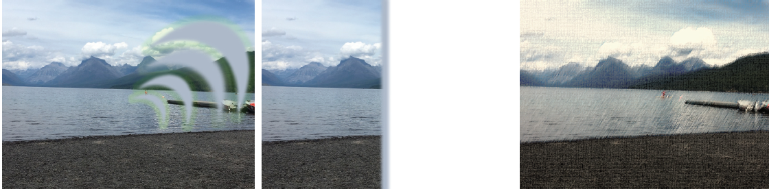
Figure. Visual disturbances seen in migraine aura. An object in the way of the patient’s view moves from center to periphery in a centrifugal, spreading pattern (left). Negative visual phenomena (center). Visual distortions or illusions of a normal scene that look fractured, underwater, or otherwise altered, often in a dynamic pattern (right).
A migraine patient may experience any single or combined version of the aforementioned visual symptoms, which completely reverse, have a stereotyped pattern for each patient, and can change over a lifetime (see Visual Snow). Visual auras can be debilitating, instilling fear in patients during highly demanding visual tasks such as driving because these auras interfere with the visual field.
Visual Snow
A persistent positive visual phenomenon associated with migraine but distinct from persistent migraine aura is known as visual snow.1 This chronic illusion persists in the absence of acute attacks and is more frequently found in patients with a history of migraine. They often describe the phenomenon as looking through TV static or snowfall.
Visual snow is distinct from persistent migraine aura (Figure). Although visual snow is harmless in isolation, any positive visual phenomenon that is new should prompt visual field testing and further workup based on accompanying historical details as described elsewhere in this article.
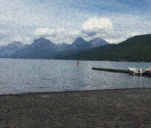
Figure. Visual snow is one type of chronic altered perception commonly reported by patients with a history of migraine. They describe a TV static-like disruption of their entire visual field that is constant and present even in the absence of headache.
1. Liu GT, Schatz NJ, Galetta SL, Volpe NJ, Skobieranda F, Kosmorsky GS. Persistent positive visual phenomena in migraine. Neurology. 1995;45(4):664-668.
DIAGNOSIS
A thorough history provides critical guidance on the differential diagnosis and workup. Neurologists frequently ask patients to keep a headache or headache aura journal or calendar. By documenting the characteristics, timing, and evolution of symptoms as well as any associated neurologic features, patients may be able to identify environmental triggers and gauge their responses to treatment more accurately. Common environmental triggers include dietary consumption of chocolate, red wine, sugar, or monosodium glutamate. High-stress environments, erratic sleep schedules, bright sunlight, strong odors, extreme exertion, and/or medication use or overuse can also be to blame. Triggers are individualized and can change over time.
The examination of a patient presenting with headache and visual disturbances should include careful documentation of BCVA and color vision, assessment for a relative afferent pupillary defect, and a dilated fundus examination. Both confrontation and formal visual field testing is highly recommended to identify optic neuropathy or homonymous defects, localizing the defect posterior to the chiasm. OCT imaging of the macular ganglion cell layer and retinal nerve fiber layer may help to localize and obtain a baseline of optic nerve structural changes, especially if optic nerve atrophy is suspected.
RED FLAGS
Several situations should raise concern about a secondary headache syndrome:
- Homonymous visual field defects;
- Loss or alteration of consciousness;
- Concurrent neurologic signs or symptoms, such as slurred speech, hemiparesis, Horner syndrome, or cranial nerve palsy. Targeted neuroimaging and a neurology workup should be performed to exclude ischemic or embolic stroke from carotid artery atherosclerosis, artery dissection, arteriovenous malformation, or intracranial aneurysm;
- Persistent vision loss or positive visual phenomena that do not fully resolve;
- Onset of migraines in patients older than 50 years of age;
- Dramatic change in the character or worsening severity of migraines or migraine aura;
- Transient monocular vision loss, often described as a curtain over vision and usually lasting only a few minutes before complete resolution. This phenomenon is indicative of amaurosis fugax and requires a retinal and cardiac workup for embolic stroke; and
- Significant associated systemic symptoms, such as scalp tenderness, jaw claudication, and arthralgias that may accompany transient vision loss or diplopia with headache. These require an emergency workup for giant cell arteritis and immediate empiric steroid treatment.
CONCLUSION
Many eye care providers are undertrained in headache classification, but knowing the basic features common among patients with associated visual disturbances and recognizing the warning signs of secondary headache are critical to making clinical decisions. Prompt referral to specialists may be necessary when the diagnosis or management strategy is uncertain.
1. Headache Classification Committee of the International Headache Society (IHS): The International Classification of Headache Disorders, 3rd edition. Cephalalgia. 2018;38(1):1-211.
2. Steiner TJ, Stovner LJ, Vos T. GBD 2015: migraine is the third cause of disability in under 50s. J Headache Pain. 2016;17(1):104.
3. GBD 2015 Neurological Disorders Collaborator Group. Global, regional, and national burden of neurological disorders during 1990-2015: a systematic analysis for the Global Burden of Disease Study 2015. Lancet Neurol. 2017;16(11):877-897.
4. Hansen JM, Charles A. Differences in treatment response between migraine with aura and migraine without aura: lessons from clinical practice and RCTs. J Headache Pain. 2019;20(1):96.