

Single-Pass Four-Throw Technique for Pupilloplasty
Narang P, Agarwal A1
Industry support: No
ABSTRACT SUMMARY
Narang and Agarwal describe their surgical technique for pupilloplasty and share their results in 27 cases. Specifically, the surgeons pass a needle once through the edges of an iris defect along the pupillary margin. Next, they perform four throws of the end of the suture through the loop in a modified Siepser slipknot technique that creates a self-retaining and self-locking helical configuration (Figure 1).
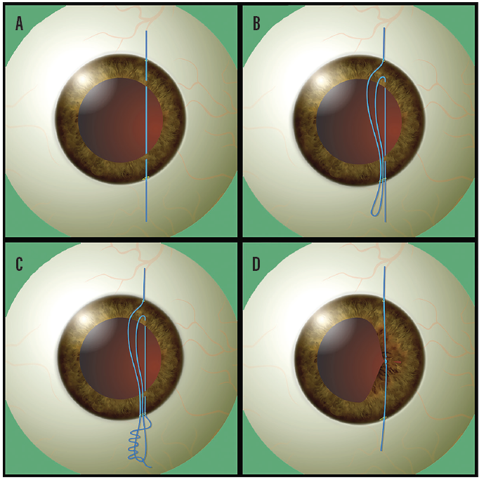
Figure 1. The needle passes through the proximal and distal portions of the iris tissue (A). The surgeon withdraws a loop of suture after approximating the proximal and distal portions of the iris tissue (B). After passing the suture through the loop (C), the surgeon pulls the suture ends, and the loop slides inside, approximating and holding the iris tissue (D).
STUDY IN BRIEF
Surgeons describe a simplified technique for pupilloplasty that requires a single pass of a suture.
WHY IT MATTERS
This technique minimizes the surgical manipulation of iris tissue, which reduces procedural time and may decrease postoperative inflammation.
The suture loop did not open in any eye during the 6-month follow-up period. The surgeons observed no iritis.
Discussion
Pinhole pupilloplasty can improve quality of vision in an eye with high corneal astigmatism and higher-order aberrations. The knot used is similar to a timber hitch. The turns of the hitch press on each other and on the iris tissue to produce self-locking units that can withstand the pull of the peripheral iris. Advantages of this pupilloplasty technique are that it is easy to learn, it is reproducible, and it requires few passes of a suture through the anterior chamber.
Application of Purkinje images for pinhole pupilloplasty and relevance to chord length mu
Narang P, Holladay J, Agarwal A, Jaganathasamy N, Kumar DA, Sivagnanam S2
Industry support: No
ABSTRACT SUMMARY
In this case series, the investigators assessed the results of pinhole pupilloplasty performed with the single-pass four-throw technique described earlier in this article. The study included eight eyes of eight patients with decreased visual acuity because of corneal higher-order aberrations (n = 7 eyes) or glare (n = 1 eye).
STUDY IN BRIEF
In a small case series, investigators used Purkinje images to guide the sizing and centration of pinhole pupilloplasty, and they evaluated the effect of the procedure on quality of vision in eyes with high astigmatism and higher-order aberrations.
WHY IT MATTERS
This study suggests a way to optimize outcomes with pinhole pupilloplasty. This technique could be an option for treating patients with high astigmatism and higher-order aberrations from a variety of causes.
Seven eyes had a posterior chamber IOL and underwent pinhole pupilloplasty alone. One eye had nuclear sclerosis and underwent the procedure in combination with phacoemulsification and IOL implantation. In each eye, the surgeon used the first Purkinje image (Purkinje P1) from the operating microscope (Lumera, Carl Zeiss Meditec) to center the pinhole pupilloplasty.
Pupilloplasty significantly reduced the mean horizontal and vertical pupillary diameters. The improvement in uncorrected distance visual acuity but not corrected distance visual acuity was statistically significant. Surgery also produced a statistically significant reduction in both mean simulated keratometry and mean chord length mu. The pupil was well centered on the Purkinje P1 images, and no major adverse events or complications occurred postoperatively.
DISCUSSION
In eyes with high astigmatism and corneal higher-order aberrations, pinhole pupilloplasty can improve quality of vision by blocking peripheral rays of light from entering the eye while funneling central and paracentral rays through the pinhole.3 Narang and colleagues used Purkinje images from a surgical microscope to guide the creation, sizing, and centration of the pupil.
Chord length mu denotes the distance between the center of the pupil and the patient-fixated coaxially sighted corneal light reflex. Pinhole pupilloplasty significantly reduced chord length mu in this study (Figure 2).
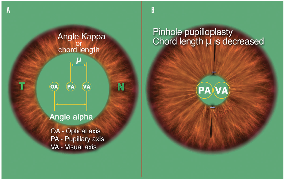
Figure 2. Chord mu is the 2D distance between the center of the pupil and the subject-fixated coaxially sighted corneal light reflex (A). A pinhole pupilloplasty reduces chord length mu (B).
The investigators attribute the observed improvement in patients’ visual acuity to the decreases in the mean size of the pupil and chord length mu. A major limitation of this study is its size. Research involving a larger number of eyes is needed to validate a correlation between improved visual acuity and reduced pupillary size and chord length mu.
1. Narang P, Agarwal A. Single-pass four-throw technique for pupilloplasty. Eur J Ophthalmol. 2017;27(4):506-508.
2. Narang P, Holladay J, Agarwal A, Jaganathasamy N, Kumar DA, Sivagnanam S. Application of Purkinje images for pinhole pupilloplasty and relevance to chord length mu. J Cataract Refract Surg. 2019;45(6):745-751.
3. Narang P, Agarwal A, Kumar DA, Agarwal A. Pinhole pupilloplasty: small-aperture optics for higher-order corneal aberrations. J Cataract Refract Surg. 2019;45(5):539-543.
