The ability to objectively diagnose patients’ quality of vision and the state of their crystalline lens is greatly aided by the measurement of ocular scatter. Objective evaluation of the health of the lens allows us to determine whether we should be performing a lens- or cornea-based procedure. Tools that allow objective evaluation of ocular scatter also generate diagnostic reports that can help to increase patients’ understanding of their pathologies and, thus, their confidence in our treatments.
I use the AcuTarget HD (AcuFocus) routinely for cataract and refractive surgery patients. The instrument uses a double-pass aberrometry approach, shining light on the retina and then measuring the transmission of this light through the tear film, cornea, and crystalline lens. The device then generates an ocular scatter index (OSI) consisting of a quantitative measurement and a qualitative analysis of the distribution pattern of scattered light in the eye. This information allows us to objectively assess visual function and determine the source of visual complaints. A high OSI may indicate corneal opacities or issues with the quality of the tear film or the crystalline lens. Conversely, a normal OSI in a patient with visual complaints could indicate issues in the retina or optic nerve.
EDUCATING PATIENTS
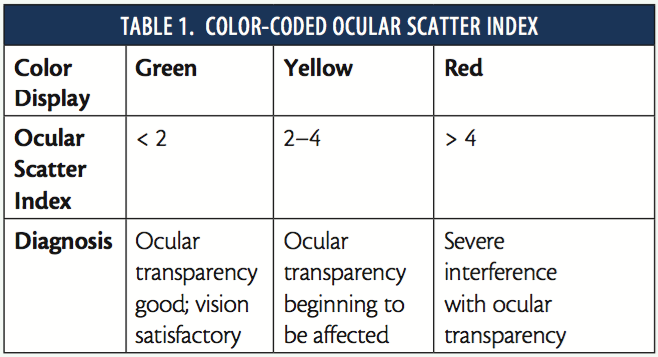
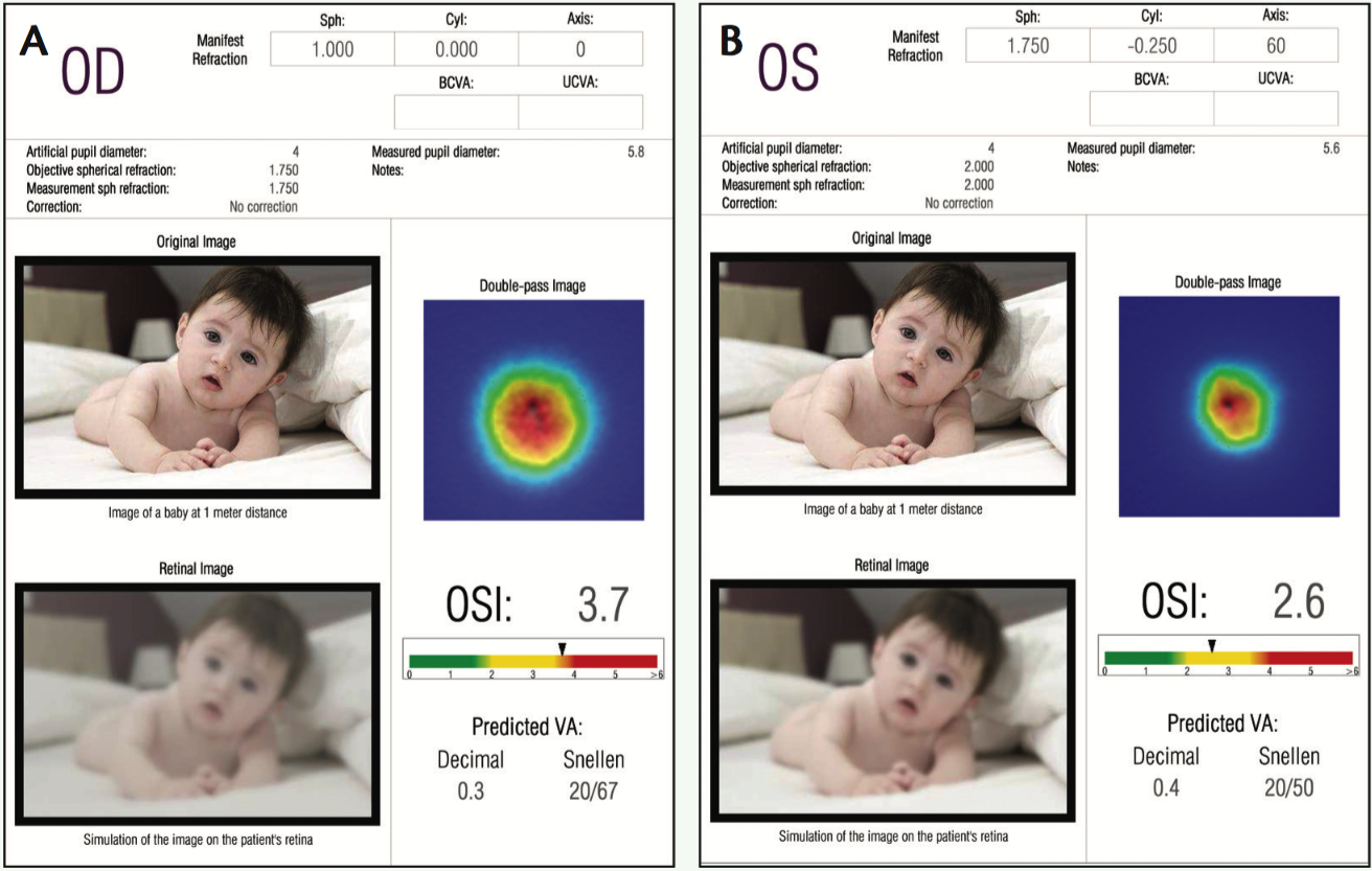
The report generated by the instrument can be shared with the patient, displaying several objective refraction images, the double-pass image, the modulation transfer function diagram, a simulated retinal image, and the OSI numbers. The OSI numbers and retinal image are particularly useful in engendering patient understanding. The numbers are illustrated on a color-coded bar that indicates increasing severity in progression from green, to yellow, to red (Table 1). The color level is tremendously useful in improving patient understanding. Patients may not understand exactly what terms such as or mean, or the significance of an OSI of 3.5, but showing them an objective number attached to a color code translates this into information they can comprehend.
The simulated image on the report represents the image that the device is sending to the retina and juxtaposes it with an image that represents what the patient is seeing. This illustrates how lens changes are affecting ocular transparency and, therefore, vision.
This type of diagnostic tool also facilitates excellent follow-up. Patients may initially present with an OSI of 1.9 or 2, which is not severe but may suggest the early presence of a cataract (Figure 1). The analyzer provides objective quantification of the patient’s subjective visual complaints and builds a base with which to compare future numbers and to aid in determining the best course of action. Detecting early cataract formation allows the surgeon to provide informed treatment and gives patients tangible evidence they can understand.
CONCLUSION
With the presentation of images and traceable numbers from the AcuTarget HD, patients can better understand the modalities of their condition and their chosen treatment. Their level of confidence in not only their care but also their physician is greatly improved when they truly understand what is happening with their vision and what is being done to address it.
Alain Saad, MD
• Anterior Segment and Refractive Surgery Department, Rothschild Ophthalmology Foundation, Paris
• Vice-director, CEROC, Paris
• dralainsaad@gmail.com
• Financial disclosure: Consultant (AcuFocus)