
The Cassini diagnostic device (i-Optics; Figure 1) offers a suite of examinations including corneal topography, mesopic and photopic pupillometry, and color photography for diagnostic purposes. The Cassini uses red, green, and yellow LEDs to perform what the manufacturer calls “true corneal shape analysis.”1 According to i-Optics, each LED is positioned in relation to its neighbors in order to give a GPS-like coordinate to each point. Patented multicolored LED point-to-point ray tracing, combined with second-Purkinje imaging technology, is used to measure the relative position of each point, with the three colors facilitating triangulation of the points (Figure 2). Elevation increases the distance between points, and depression decreases the distance. These principles allow accurate and repeatable measurements of total corneal astigmatism.
The Cassini does not use edge detection in its algorithms, as does conventional Placido-disc–based topography. Therefore, smeared or double-reflected LEDs cannot skew results. This allows the device to image all corneas, even highly irregular ones.
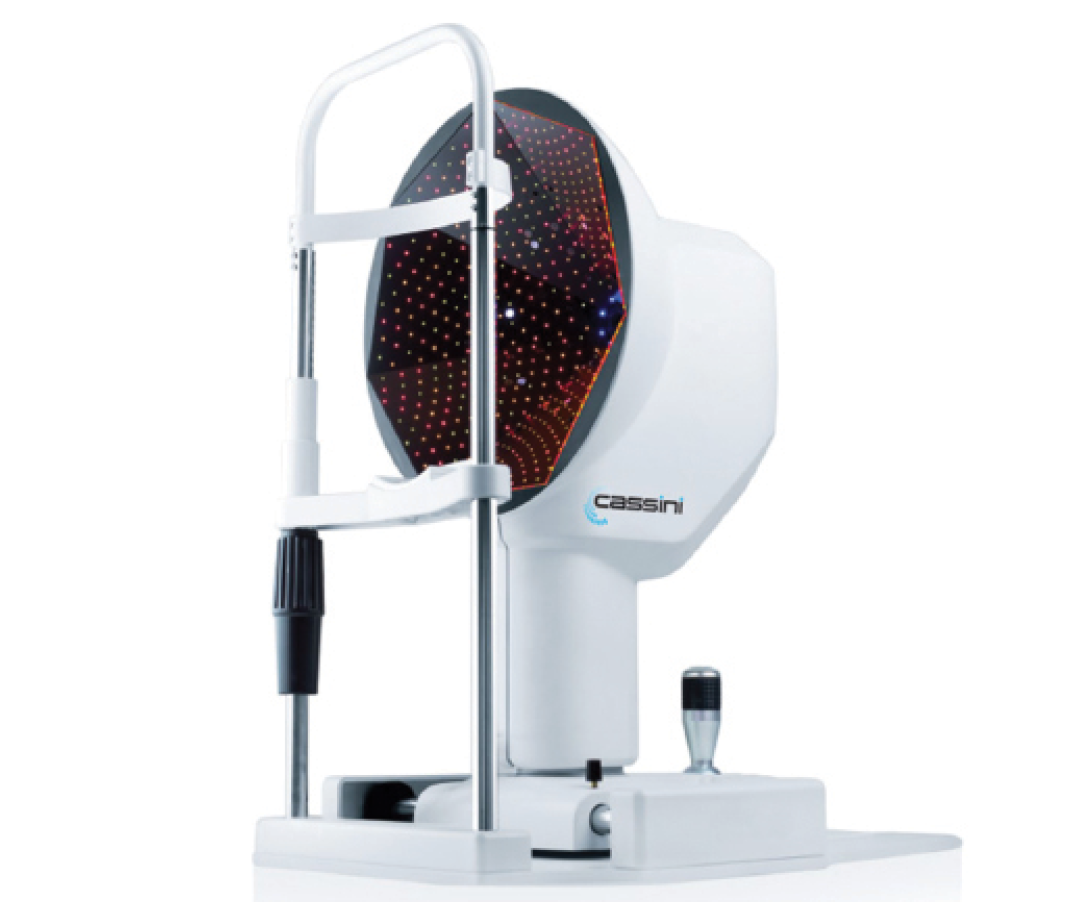
Figure 1. The Cassini diagnostic device offers a suite of examinations including corneal topography, mesopic and photopic pupillometry, and color photography for diagnostic purposes.
The technology particularly supports toric IOL implantation, providing information to select the power and proper positioning of toric IOLs. The device repeatably measures the axis of cylinder to an accuracy within 3° (as compared with 13° with Placido technology) and the magnitude to within clinical margins (2%), according to the company. Data capture is instantaneous, with submicron accuracy provided by the color LED triangulation technology. Alignment for data capture is intuitive, with color changes indicating the degree of correct alignment. Each image is stamped with the quality factor of the capture.
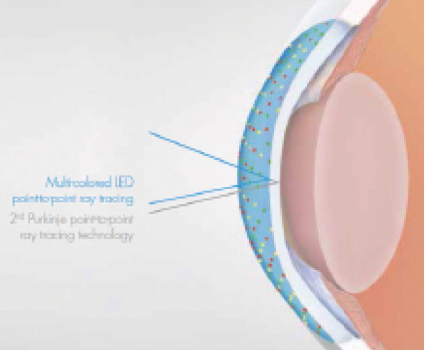
Figure 2. Patented multicolored LED point-to-point ray tracing provides a GPS-like analysis of the cornea.
According to the company, the Cassini addresses the four most common problems associated with astigmatism correction: (1) surgically induced astigmatism, addressed by its accurate postoperative measurements; (2) preoperative measurement errors, addressed by its accuracy and precision, (3) correct toric IOL alignment, addressed by its highly accurate marking capabilities, and (4) marking errors, addressed by its real-time eye-tracking systems.
With the Cassini technology, there is no central corneal scotoma. The multicolored LED coverage is uniform across the entire cornea.
The information screen displays all relevant data, including the captured image with axis details, corneal elevation maps, tangential and axial curvatures, corneal refractive power, and an overview of Zernike terms, in addition to measurement of steep and flat keratometry readings and astigmatism. Any two exams can be compared differentially. Measurable keratoconus indices include surface asymmetry and surface regularity.
The system’s topographic map displays include axial, refractive, tangential, elevation, and higher-order aberrations, and its topographic indices include shape factor, eccentricity, asphericity, and form factor. The device can connect to femtosecond lasers, operative guidance systems, and toric IOL calculators. A patient management program is incorporated. n
1. Cassini True Technology. i-Optics website. http://i-optics.com/us/products/cassini-2/true-technology-2/. Accessed June 22, 2015.
Lensar to Integrate Wirelessly With the Cassini
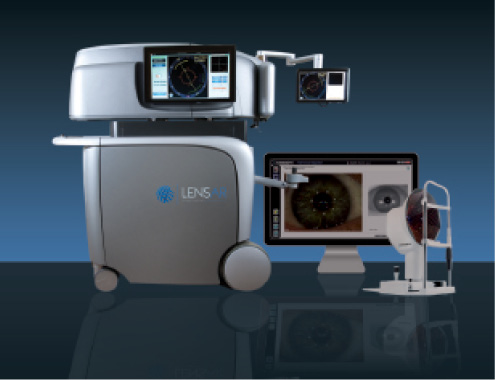
Figure 1. The Lensar Laser System now integrates wirelessly with the Cassini.
