

What is your overall impression of the Cassini?
I have had the pleasure of working with the Cassini Corneal Analyzer (i-Optics) for more than 2 years. At first glance, it looks like any other topographer; however, when you see it in action and evaluate the performance, it is clear that the Cassini is indeed a different breed. Trichromatic LEDs provide a detailed depiction of the anterior corneal surface that was not possible before with other devices, and, as a result, I have changed how I approach the corneal maps I traditionally use for assessing corneal shape. I tend to scrutinize the axial maps of other elevation-based devices—for instance, the Placido-ring display to evaluate a dry eye or the curvature maps for irregular corneas. The multipoint discrimination of the Cassini enables a more accurate and nearly complete representation of the anterior surface elevation and curvature, and its posterior corneal analysis provides information on total corneal power.
How do you use the Cassini in clinical practice?
The Cassini delivers the preoperative data for my refractive cataract platform. In conjunction with the Streamline upgrades to the Lensar Laser System (Lensar), the corneal refractive information is seamlessly and efficiently fed to the laser for arcuate incision planning and reproducible clear corneal incision placement. (Editor's Note: See Lensar to Integrate Wirelessly With Cassini for more information on the Streamline upgrades.) These features help to standardize my surgically induced astigmatism.
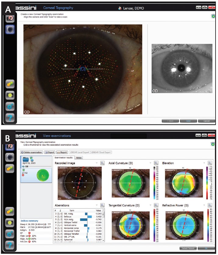
Figure 1. Capture screen of the Cassini (A). View of the corneal topography examination (B).
Furthermore, I have grown particularly fond of the Cassini Corneal Shape Analyzer for surgical management of corneal disease. For patients with corneal ectasia, preoperative planning for intrastromal corneal ring segments and intraoperative guidance for conductive keratoplasty is driven by the Cassini system with remarkable precision.
What are the advantages of the Cassini compared with other ocular biometry technologies?
The distinct advantage is the enhanced assessment of the anterior corneal shape that is provided with the Cassini analyzer (Figure 1). The pseudorandomized color LEDs are superior to traditional monochromatic Placido rings for detailed interpretation of the anterior corneal curvature. For example, evaluation of an irregular, asymmetric cornea is frequently limited and a source for confusion with a traditional Placido device, and poor extrapolation with Scheimpflug tomography is frequently depicted as data dropout. By comparison, using the Cassini Corneal Shaper Analyzer, the indices over the focal areas of irregularity are preserved. In my clinic, the Cassini provides a wealth of knowledge regarding front and back corneal surfaces across the full spectrum of patient presentations.
Jonathan D. Solomon, MD
• Surgical/Refractive Director of Solomon Eye Associates, Bowie, Maryland
• jdsolomon@hotmail.com
• Financial disclosure: Consultant (i-Optics, TrueVision)
