
Rapid measurement is vital for patient comfort and efficient workflow. The AL-Scan Optical Biometer (Nidek; Figure 1) measures six values for cataract surgery in 10 seconds: axial length, anterior chamber depth, central corneal thickness, white-to-white distance, pupil size, and corneal curvature radius, according to the company.1 The device incorporates Nidek’s 3-D auto-tracking and auto-shot technologies, ensuring ease of operation and comfort for the user. The 3-D auto-tracker follows eye movements in the x, y, and z planes; once correct alignment is achieved, the auto-shot feature automatically captures the image and data.
In cataractous eyes, the advanced algorithms incorporated in this diagnostic device help to filter signal from noise, boosting strength of the signal output in dense cataracts. In extremely dense cataracts that are not conducive to optical biometry, an optional built-in ultrasound biometer is available for the AL-Scan. With this feature, virtually any eye can be measured without having to move the patient or connect to an external ultrasound device.
The software of the AL-Scan includes nine IOL power calculation formulas, including Regression and Regression II, Binkhorst, Hoffer Q, Holladay, Haigis, Camellin-Calossi, and Shammas PL. Once the measurement capture is completed, IOL power is automatically calculated using the acquired data. Surgeons can improve accuracy by using the unit’s IOL A-constant optimization feature; the AL-Scan statistically calculates optimum A-constants based on postoperative refraction data.
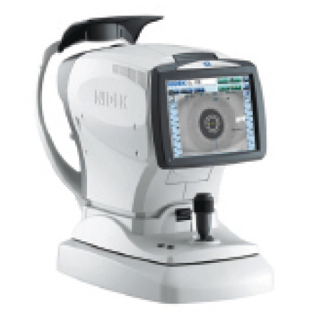
Figure 1. Measuring six values in 10 seconds, the AL-Scan incorporates 3-D auto-tracking and auto-shot technologies.
The AL-Scan also provides assistance for toric IOL implantation. On the acquired frontal image of the cornea, iris, and conjunctiva, the device can draw a line passing through a vessel or other landmark to indicate the angle from the steepest corneal meridian.
The line and angle are clearly marked and overlaid onto the eye image, which can be taken to the operating room to act as a guide for toric IOL implantation.
In addition to the frontal image, the AL-Scan supplies other anterior segment views, including cross-sectional lens image, pupil image, and reflected image of corneal mires for astigmatism assessment. The display is provided on a tiltable 8.4-inch color LCD touchscreen. n
1. Optical Biometer AL-Scan. Nidek Co. Ltd. website. http://www.nidek-intl.com/product/ophthaloptom/diagnostic/dia_cornea/al-scan.html. Accessed June 15, 2015.
