
The Refractive Power/Corneal Analyzer OPD-Scan III (Nidek; Figure 1) is a third-generation combined aberrometer and corneal topographer that is faster, more accurate, and more user-friendly than its predecessor, the OPD-Scan II, according to the manufacturer.1 The combination of aberrometry and topography in one unit offers eye care practitioners versatility and provides both broad and precise information about the eye that allows comprehensive analysis and assessment, according to company literature.
The device is a five-in-one workstation, combining wavefront aberrometry, corneal topography, autokeratometry (for keratometry [K] readings), autorefractometry, and pupillometry. The wavefront aberrometer provides assessment of visual acuity and quality of vision that goes beyond standard refraction and keratometric data. The 9.5-mm diameter wavefront ensures complete coverage of almost any pupil. Data are gathered from 2,520 points, 175% the number of points measured by the OPD-Scan II, thereby significantly increasing measurement accuracy and spatial resolution, according to the company. Information from the aberrometer can be used to create simulated retinal contrast acuity sensitivity and visual charts that provide objective quantification of visual clarity.
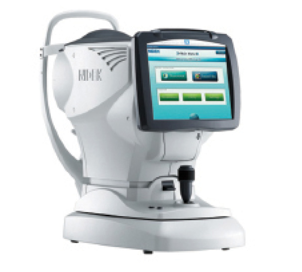
Figure 1. The OPD-Scan III combines wavefront aberrometry, corneal topography, autokeratometry (for K readings), autorefractometry, and pupillometry.
The topographer generates intuitive maps of the corneal surface and indices of corneal pathology such as keratoconus or pellucid marginal degeneration. With 33 blue Placido mires providing a minimum of 11,880 data points, the resolution is increased in comparison with the OPD-Scan II. The blue mires allow greater precision in ring detection, and the reduced level of illumination presents a more comfortable experience to the patient.
The autorefractometer provides accurate refractions under mesopic and photopic conditions—data that may be important for planning refractive surgery or analyzing common refractive problems. The autokeratometer, in addition to standard keratometric measures such as simulated K, also provides novel corneal surface descriptors, such as average pupil power and effective central corneal power, which can aid in calculating IOL power for eyes after refractive surgery. Finally, the pupillometer/pupillographer measures photopic and mesopic pupil size and documents the shape of the pupil under these conditions, which may alter refraction and affect surgical planning.
The overview summary page displays multiple maps, indices, and images, and it serves the surgeon as a guide for clinical decision-making. The information displayed includes an irregularity index, with higher-order aberrations divided into corneal, internal, and total, to help determine the best strategy for vision correction. A separate display of corneal spherical aberration aids in the selection of the best aspheric IOL. Color-coded classification indices help to identify eyes that have undergone corneal refractive surgery, and the astigmatism index helps with the planning of toric IOL implantation, including incision placement and lens alignment.
In addition to the overview summary, a number of more focused summary screens, customizable to the surgeon’s preferences, can be called up to assist with particular tasks. These include cataract, toric IOL, optical quality, and white-to-white summaries. Pages with multiple maps for comparison can also be called up. n
1. Refractive Power/Corneal Analyzer OPD-Scan III. Nidek website. http://www.nidek-intl.com/product/ophthaloptom/diagnostic/dia_cornea/opd-scan3.html. Accessed June 23, 2015.
