
Myopia is characterized by a mismatch between the axial length of an eye and the refractive power of the eye's optics. Degenerative myopia is characterized by an axial length of greater than 26.5 mm and a progressive retinal and choroidal atrophy compromising retinal function. Myopia is endemic among young adults in certain areas of Asia, with up to 90% of those of graduation age affected.1 In Japan, progressive myopia represents the third leading cause of blindness.2,3 Hypotheses for underlying mechanisms of myopia include form deprivation and forced near work. Therapeutic approaches include increased exposure to natural environmental light to overcome dopamine suppression and suppression of accommodation.
In an attempt to arrest axial growth mechanically, several scleral reinforcement techniques have been proposed using either human donor sclera or synthetic materials. One problem with the approaches that have been proposed thus far has been that buckling surgery in these eyes comes with an increased risk of complications such as perforation, thrombosis, and ischemia.
At a Glance
• The concept of scleral CXL may someday lead to a
therapeutic approach to arrest scleral elongation
and progressive myopia; proof of concept has been
demonstrated in vivo and ex vivo.
• The combination of light and a chromophore may not
be ideal for performance of scleral CXL.
• It remains to be seen which CXL technique will prevail
for the sclera: photoactivation of a chromophore,
chemically induced CXL, or other approaches that have
yet to emerge.
SCLERAL REINFORCEMENT WITH CXL
The concept of scleral reinforcement with CXL using a chromophore and light is a fascinating one. It would enable the surgeon to increase scleral stiffness without the need for complex posterior pole surgery.
A direct relationship between the severity of form-deprivation myopia and collagen crosslinking was described in 1994 by McBrien and Norton.4 Applying a chemical crosslinking blocker, i-aminoproprionitrile (APN), in a tree shrew model of form-deprivation myopia in vivo, they showed that APN-treated form-deprived eyes developed significantly lower degrees of myopia than control animals receiving saline and form-deprivation.
In 2004 and 2005, Wollensak and colleagues investigated the effects of riboflavin UV-A CXL on the human sclera ex vivo and found significant increases in scleral biomechanics after treatment.5,6 Four years later, Wollensak7 demonstrated an in vivo effect on rabbit scleras using 370 nm and a standard fluence of 5.4 J/cm2. More recently, Iseli and colleagues have shown that similar results may be achieved using visible blue light at 450 ±50 µm.8
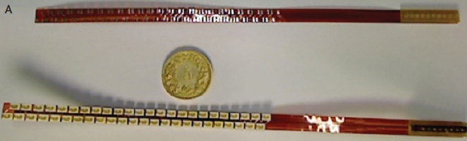
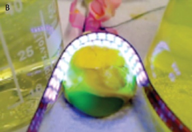
Figure 1. Printed circuit board with and without light-emitting diodes (A). The sclera in this porcine eye is saturated with riboflavin, and the printed circuit board is wrapped around the posterior pole (B).
LIGHT RIGHT FOR SCLERAL CXL?
The concept of using light and riboflavin for CXL, which worked so well in the cornea, might not be ideal for the sclera, however. The corneal CXL concept may not be simply translated to the sclera without modification for two reasons.
Reason No. 1: UV-A light and even visible blue light alone and riboflavin alone may be potentially toxic to the underlying choroid and outer retina, and little is known about the effect of photoactivated riboflavin on the outer retina. Along with colleagues, one of us (FH), in the early 1990s, showed that short wavelength light, when applied excessively, can irreversibly damage the retinal photoreceptors by apoptosis.9,10
The light-transmission characteristics of the sclera are distinctly different from those of the cornea, and the wavelength of 365 nm used in corneal CXL might not be ideal for the sclera. For scleral CXL, Zhan et al showed that 50 minutes of UV-A exposure, with a wavelength of 365 nm applied at 3 mW/cm2 (total enegry dose 7.2 J/cm2), led to distinct retinal damage in the outer retina.11
Reason No. 2: Miniaturization of a scleral CXL device is challenging due to technical constraints. In other words, how would the light be delivered to the posterior part of the globe? Whatever device is used must emit homogeneous and high-powered UV-A and visible blue light. The use of optical fibers is not possible because the size of the fiber is proportional to the amount of UV-A power transported, with important binding limitation. Light-emitting diodes (LEDs) are small in size, but they provide inhomogeneous light distribution and limit the flexibility of the device. LEDs also generate heat, which might potentially harm the retina and sclera.
PROGRESS MADE
The ELZA Laboratory of Ocular Sciences has developed a relatively small flexible printed circuit board (5 mm in length and width and 1.2 mm high) with small UV-A 365-nm LEDs soldered onto it (Figure 1A). This device has been used to perform scleral CXL in ex vivo porcine eyes (Figure 1B).
However, light might not be the way to go. An interesting alternative to light-mediated CXL may be the use of chemical CXL with, for example, genipin or nitroalcohols.12,13
CONCLUSION
Scleral CXL stands today where corneal CXL was in the late 90s: at the very beginning, representing a fascinating concept that may someday lead to a therapeutic approach for progressive myopia. However, the settings and parameters known from UV-A and riboflavin corneal CXL cannot simply be transferred to the sclera.
It remains to be seen which CXL technique will prevail for the sclera: photoactivation of a chromophore, chemically induced CXL, or other approaches that have yet to emerge. n
1. Morgan IG, Ohno-Matsui K, Saw SM. Myopia. Lancet. 2012;379(9827):1739-1748.
2. Iwase A, Araie M, Tomidokoro A, et al. Prevalence and causes of low vision and blindness in a Japanese adult population: the Tajimi Study. Ophthalmology. 2006;113(8):1354-1362.
3. Yamada M, Hiratsuka Y, Roberts CB, et al. Prevalence of visual impairment in the adult Japanese population by cause and severity and future projections. Ophthalmic Epidemiol. 2010;17(1):50-57.
4. McBrien NA, Norton TT. Prevention of collagen crosslinking increases form-deprivation myopia in tree shrew. Exp Eye Res. 1994;59(4):475-486.
5. Wollensak G, Spoerl E. Collagen crosslinking of human and porcine sclera. J Cataract Refract Surg. 2004;30(3):689-695.
6. Wollensak G, Iomdina E, Dittert DD, et al. Cross-linking of scleral collagen in the rabbit using riboflavin and UVA. Acta Ophthalmol Scand. 2005;83(4):477-482.
7. Wollensak G, Iomdina E. Long-term biomechanical properties of rabbit sclera after collagen crosslinking using riboflavin and ultraviolet A (UVA). Acta Ophthalmol. 2009;87(2):193-198.
8. Iseli HP, Korber N, Karl A, et al. Damage threshold in adult rabbit eyes after scleral cross-linking by riboflavin/blue light application. Exp Eye Res. 2015;139:37-47.
9. Hafezi F, Marti A, Munz K, et al. Light-induced apoptosis: differential timing in the retina and pigment epithelium. Exp Eye Res. 1997;64(6):963-970.
10. Hafezi F, Steinbach JP, Marti A, et al. The absence of c-fos prevents light-induced apoptotic cell death of photoreceptors in retinal degeneration in vivo. Nat Med. 1997;3(3):346-349.
11. Zhang Y, Zou C, Liu L, et al. Effect of irradiation time on riboflavin-ultraviolet-A collagen crosslinking in rabbit sclera. J Cataract Refract Surg. 2013;39(8):1184-1189.
12. Paik DC, Saito LY, Sugirtharaj DD, et al. Nitrite-induced cross-linking alters remodeling and mechanical properties of collagenous engineered tissues. Connect Tissue Res. 2006;47(3):163-176.
13. Liu TX, Wang Z. Collagen crosslinking of porcine sclera using genipin. Acta Ophthalmol. 2013;91(4):e253-257.
Farhad Hafezi, MD, PhD
• Chief Medical Officer, The ELZA Institute, Dietikon/Zürich,
Switzerland
• Professor of Ophthalmology, Faculty of Medicine, University of
Geneva, Switzerland
• Clinical Professor of Ophthalmology, University of Southern
California, Los Angeles
• info@elza-institute.com
• Financial disclosure: Co-inventor of CH880-B941 application
Olivier Richoz, MD, PhD
• Cornea and Refractive Fellow, Royal Victoria Hospital, Belfast,
United Kingdom
• richozolivier@gmail.com
• Financial disclosure: Co-inventor of CH880-B941 application
