No. 1: First Experience with Verus
By Aaron Waite, MD
Dr. Waite shows his first experience with the Verus ophthalmic caliper (Mile High Ophthalmics) and outlines seven steps for successful use of the device. The Verus, a biocompatible silicone ring designed to enhance the accuracy and reproducibility of the continuous curvilinear capsulorrhexis, fits into the standard cataract procedure flow and results in a well-centered and round 5-mm capsulotomy.
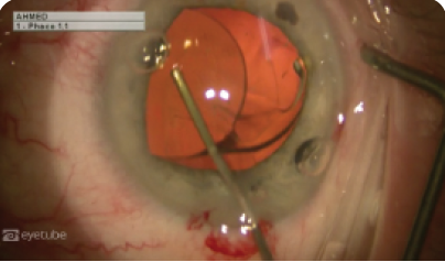
No. 2: Cataract Surgery Using a Vitreous Cutter Without Phacoemulsification
By Gurkan Erdogan, MD
Dr. Erdogan uses a vitreous cutter to remove a cataract in an uncomplicated case.
No. 3: Surgeon’s Hand Position During Cataract Surgery: Pearls for Residents
By Howard Gimbel, MD
Hand and arm positions during cataract and refractive surgery are not often visible when sharing surgical expertise via video or a satellite broadcast of live surgery. Dr. Gimbel presents insights for hand techniques that have been successful for cataract and refractive surgery.
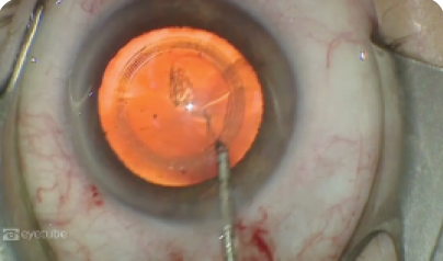
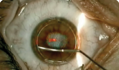
No. 4: IOL Exchange and Double Optic Capture for the Management of Uveitis-Glaucoma-Hyphema Syndrome
By Xavier Campos, MD; Iqbal Ike K. Ahmed, MD; and Manjool Shah, MD
The surgeons demonstrate removal of a one-piece acrylic IOL in a bag-sulcus position in a patient with symptomatic uveitis-glaucoma-hyphema syndrome. A posterior continuous capsulorrhexis is created, and a three-piece IOL is subsequently implanted in the sulcus and captured posteriorly through the anterior and posterior capsulotomies.

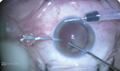
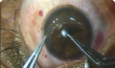
No. 5: Iridodialysis, Capsular Bag Repair, Phacoemulsification, and IOL Insertion
By Allon Barsam, MB, BS, MA, FRCOphth
Dr. Barsam shares a challenging case involving a patient who had experienced blunt trauma, resulting in a large iridodialysis and more than 120º of zonular weakness. He presents a step-by-step approach to the management of cataract surgery with multiple ocular traumas.
No. 1: Longest Refractive Day
By Soosan Jacob, MS, FRCS, DNB
Dr. Jacob shows the sequence of events and explains the management strategy that she employed to manage a buttonhole. Her efforts eventually led to a satisfied patient.
No. 2: Disposable Instruments for Creating an Oval Flap
By Asim Piracha, MD
Dr. Piracha creates an oval flap with the 150-kHz iFS femtosecond laser (Abbott Medical Optics) and uses disposable, single-use instruments, including an adjustable speculum, Sinskey hook, and LASIK cannula (Moria) throughout the procedure.
No. 3: ReLEx SMILE Technique
By Joaquín Fernández, MD
Dr. Fernández performs ReLEx small incision lenticule extraction for myopia and astigmatism correction. The video is narrated by Almudena Valero Marcos, MD.
No. 4: FILI Keratoconus
By Sri Ganesh, MBBS, MS, DNB
Dr. Ganesh performs femtosecond intrastromal lenticule implantation combined with accelerated CXL for the treatment of mild to moderate keratoconus. During the procedure, a donut-shaped lenticule is placed in a corneal pocket in order to improve corneal shape and thickness and to reduce aberrations.
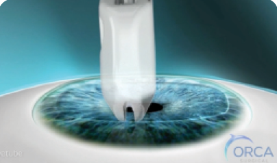
No. 5: EBK ProcedureBy ORCA Surgical
This video demonstrates the EBK technique for epithelium removal. EBK preserves the Bowman layer and creates clear and graduated borders for enhanced healing. n

