Lens disassembly is a crucial step of cataract surgery. When the phaco tip is within the main cataract incision, typically either the tip is pushed into the lens material or high vacuum is initiated to attract the lens. Both maneuvers can cause problems. Pushing the tip into the lens can damage the zonules, and high vacuum can accidentally grab important structures such as the iris and posterior capsule.
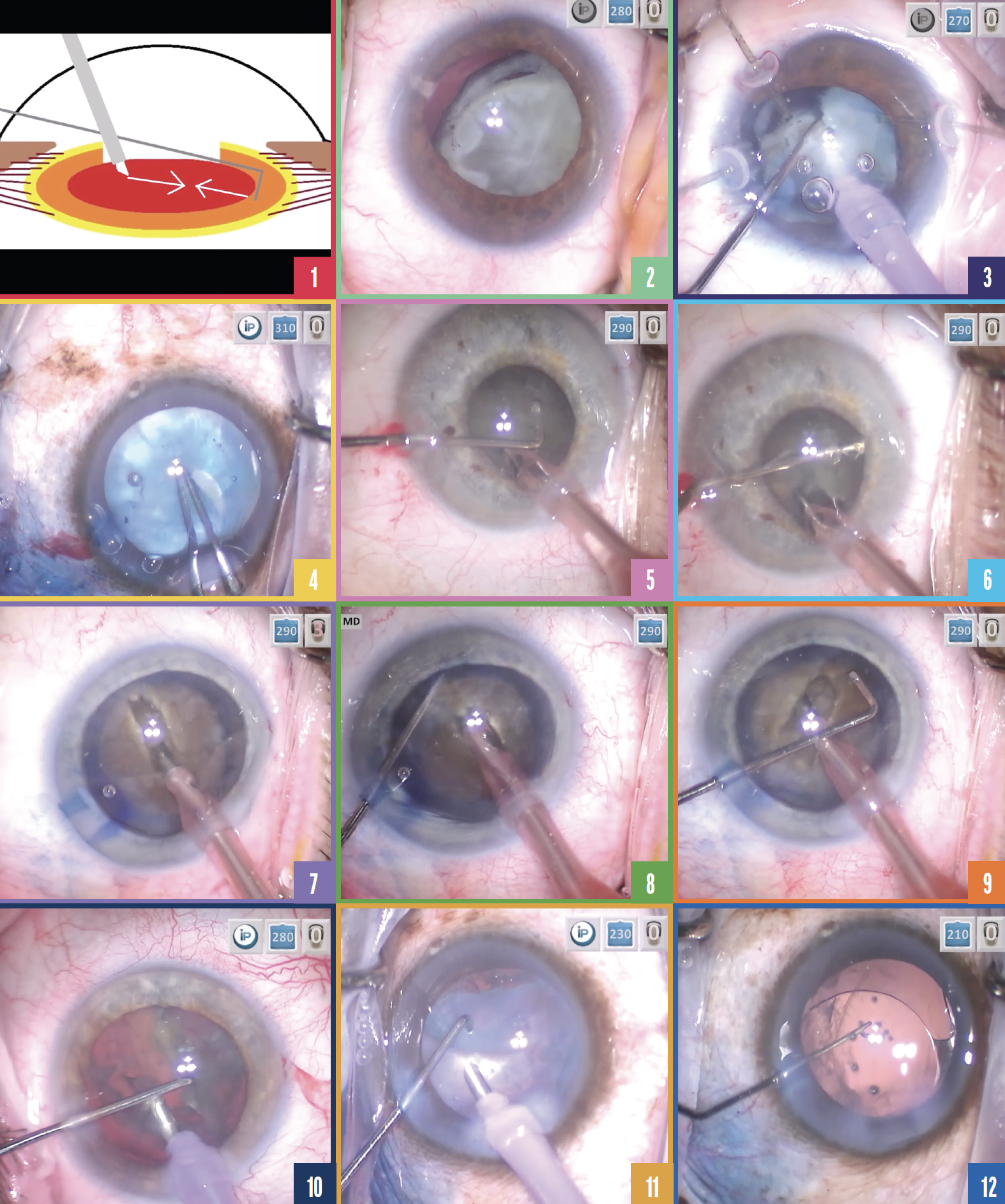
An alternative is to place the chopper around the lens equator across from the phaco tip. This allows the opposing forces between the instruments to disassemble the lens (Figure 1).

Figure 1. Placing a chopper around the lens equator across from the phaco tip allows the lens to be disassembled by the opposing forces (arrows) of the two instruments.
Figure 2. Ectopia lentis.
Figure 3. After the placement of capsule retractors and a capsular tension ring, opposing mechanical forces alone are used to divide a dislocated crystalline lens in half.
Figure 4. The anterior capsule is punctured in an eye with an intumescent cataract.
Figure 5. The lens is divided by successive applications of opposing forces.
Figure 6. A chopper is placed around the right hemi-nucleus in an eye with a small pupil.
Figure 7. A hypermature lens wobbles during attempts at sculpting. Cutting is inefficient.
Figure 8. The chopper provides a counterforce to the phaco tip, which can now fracture the hypermature lens.
Figure 9. The crack does not extend across the nucleus, so the chopper is positioned around the other side of the lens equator and pulled toward the phaco tip, which splits the nucleus.
Figure 10. The hemi-nucleus will not spin. A chopper is placed under the edge of the capsulorhexis.
Figure 11. The chopper is used to pull thickened epinuclear material centrally.
Figure 12. The IOL haptics are carefully oriented perpendicular to the tear in the anterior capsule.
FIVE SITUATIONS
The technique can be particularly helpful in five situations.
No. 1: Ectopia lentis. The zonules are weak and unstable in an eye with ectopia lentis (Figure 2). Force applied to the capsular bag or lens can further damage the zonules. The risk of rupturing the posterior capsule therefore increases. Opposing forces used to crush the lens are directed toward the center, away from the zonules and capsular bag, thereby increasing the safety of nuclear disassembly.
In the video (see video below), capsule retractors and a capsular tension ring are placed. Successive maneuvers in which the chopper is drawn toward the phaco tip divide the nucleus in half. No ultrasound energy or vacuum is required (Figure 3).
No. 2: Intumescent lens. After the anterior capsule is punctured in an intumescent lens, there is a risk that the tear will extend radially, potentially leading to a dropped nucleus. By placing the chopper in opposition to the phaco tip, the opposing mechanical forces can be applied in a controlled manner without placing traction on the capsular bag or stress on the anterior capsule. As a result, there is no reason for the capsular tear to propagate posteriorly.
In the video, the anterior capsule is punctured, and the Argentinian flag sign becomes visible (Figure 4). The chopper is placed around the lens equator, and the successive application of opposing and centrally directed forces divides the lens in half (Figure 5). Both the chopper and the phaco tip are used to chop the nucleus, allowing controlled disassembly of the lens without the use of ultrasound energy or vacuum and reducing the risk for a capsulorhexis runout.
No. 3: Small pupil. The challenge posed by an eye with a small pupil is poor visibility. An alternative to the placement of pupillary expansion devices is to hook the peripheral lens with the chopper, tilt the phaco tip vertically and subincisionally, and fracture the lens by drawing the chopper and phaco tip toward the center to crush the lens. The chopper is then positioned around the right hemi-nucleus and pulled centrally toward the phaco tip, further dividing the lens (Figure 6). The key to the success of this maneuver is for the instruments to flank the nucleus or nuclear fragment on both sides.
No. 4: Hypermature lens. Sometimes, a lens is too dense to cut. Attempts to sculpt put stress on the zonules, which may already be weak. Placing the chopper around the lens equator holds the lens from the other side. Now when the phaco tip pushes down on the lens, it presses against the chopper instead of the zonules. This increases safety as well as sculpting efficiency.
In the video, wobbling of the lens makes sculpting inefficient (Figure 7). Positioning the chopper around the lens equator and hugging it from the periphery provides counterforce to the lens. It can then be fractured from the terminal side and the tear propagated all the way through (Figure 8), preventing a posterior plate phenomenon. The crack, however, does not extend across the nucleus. The chopper is therefore positioned around the other side of the lens equator and pulled toward the phaco tip, which easily divides the nucleus (Figure 9).
No. 5: Fragment removal. Using a chopper to support the lens at the level of the equator allows the lens to be pulled up from the equatorial side and out of the bag. Rather than attempt to attract pieces with vacuum, which may lead to grabbing the posterior capsule by mistake, lens material is hooked with the chopper from the equator and extracted from the bag.
In the video, the nucleus will not spin. The chopper is passed underneath the edge of the capsulorhexis (Figure 10). The nucleus is hooked and rotated up and out of the bag at an angle. It is then split in half.
A BONUS USE
No. 6: Thick epinucleus. The opposing forces maneuver can also facilitate removal of thick epinucleus. In the video, an attempt to grab the material with vacuum fails. The opening in the anterior capsule is discontinuous, and the capsular bag is floppy. Further use of vacuum or the application of ultrasound energy would be risky. Instead, a chopper is placed around the equator and used to pull the epinucleus centrally (Figure 11).
The movement is gentle. The instrument touches neither the capsular bag nor the tear in the anterior capsule. The epinucleus is then removed with minimal vacuum. The IOL is implanted, and the haptics are oriented perpendicular to the anterior capsular tear (Figure 12). The OVD is removed, and surgery is concluded.
CONCLUSION
All of the steps of cataract surgery affect the final outcome, but I find being able to place the chopper around the lens equator confidently and safely is my favorite timeless tip.


