


Despite recent advances in the treatment of ocular surface disease (OSD), new tools are needed to improve our management of this complex group of conditions. The purposes are threefold: to better detect, to better treat, and to better understand these conditions and how they affect our patients—not just in the clinic, but also in the real world. This is especially important in patients who develop symptoms of dry eye disease (DED)—a common and important subset of OSD—after refractive surgery.
CURRENT NEEDS IN OSD MANAGEMENT
Eye care providers must be able to accurately assess the impact of OSD on each patient’s quality of vision and quality of life in order to tailor daily care accordingly. We must also be able to objectively quantify the severity of the disease; in order to do this, we need an OSD index that reflects the severity and scope of the disease in a quantitative and objective manner. This also requires the quest for pathologic processes involving previously unrecognized pathways of OSD that may enable earlier diagnosis and identification of new therapeutic targets.
AT A GLANCE
- Despite recent advances in the treatment of OSD, new tools are needed to improve the management of this complex group of conditions.
- Physicians should strive to understand the effects that DED has on patients, not just in the clinic, but in the real world, and adapt their care accordingly.
- Efforts are being made to find better markers to help standardize the diagnosis and grading of DED.
Further, we need criteria that allow us to analyze the results of OSD research, particularly multicenter studies, and to validate the conclusions of this research. Regulatory authorities require clinical proof to validate new treatments.
ASSESSING IMPACT ON QUALITY OF VISION AND QUALITY OF LIFE
For years, DED has been recognized as a risk factor for depression, with psychological and social impacts on a large population of affected patients. Thus, dedicated questionnaires about quality of life and vision-related quality of life, as well as other testing methods, have been developed to help elucidate the consequences of DED on patients’ daily living.
Questionnaires. The Ocular Surface Disease Index (OSDI) questionnaire aims to quantify the severity of DED by evaluating a patient’s symptoms, assessing the condition’s impact on his or her daily activities, and identifying environmental factors that may be contributing to it. The Impact of Dry Eye on Daily Life (IDEEL) questionnaire focuses specifically on DED’s impact on the patient’s daily activities, emotional health, and work in order to assess the consequences of the disease.
Studies validating the OSDI and IDEEL questionnaires have confirmed that these instruments provide accurate assessments of the impact of DED on patients’ daily lives.1,2 The effects of DED are especially prominent in today’s world, in which common activities include reading, computer and mobile device use, and driving.
Driving simulator. A few years ago, my colleagues and I developed a driving simulator to assess the visual performance of patients with DED (Figure 1).3 During simulations, patients were asked to click a button to identify designated targets. We found that there was a significant increase in response time to identify targets in patients with DED compared with healthy individuals. More specifically, we observed that, at crossroads or roundabouts, there was a tremendous increase in response time among DED patients.
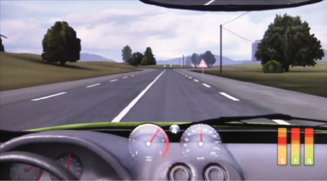
Figure 1. In a driving simulation, patients with DED had a slower response time in identifying targets compared with healthy individuals.3
Our findings indicated that the more a patient with DED needs to concentrate, the poorer his or her visual abilities become. Interestingly, it may be that patients’ response time correlated with their quality of life, suggesting that this measure could, in part, at least reflect the severity of the disease. As mentioned previously, it is crucial that we develop OSD indices that better reflect the entire severity of the disease in an objective and quantitative way. The development of a visual or optical index would help us to better define the social impact of DED.
Contrast sensitivity. Contrast sensitivity is known to be increased in patients with DED, mainly at high spatial frequencies. Standard tests for contrast sensitivity, however, are static, subjective, complex, time-consuming, and not very specific.
Dynamic optics. Thanks to refractive surgery, we have seen the development of instruments that can measure dynamic optics, including videotopography, interferometry, double-pass aberrometry, and dynamic aberrometry. These tools are now helping us to better understand and evaluate the impact of DED on optical quality.
Double-pass aberrometry with the Optical Quality Analysis System (Visiometrics) has identified an increase in ocular surface index after blinking in DED patients compared with healthy individuals. This is one objective way to measure deviations in optical quality. However, this test is poorly sensitive and not correlative with clinical signs.
In 2012, we used dynamic aberrometry on the KR-1W aberrometer (Topcon) to quantify the deviation and optical quality of eyes with DED.6 With serial measurements of ocular and corneal higher-order aberrations (HOAs) for 10 seconds after blinking, we found that patients with DED demonstrated a degradation in point spread function while controls did not. Based on our findings, we defined what we call the progression index (PI), a change in total ocular HOAs, total corneal HOAs, and corneal third-order aberrations. After blink, this PI was higher in patients with DED than in healthy individuals. We determined this progression index to be objective, quantitative, and sensitive.
The PI of HOAs is positively correlated with clinical signs such as tear film breakup time, symptoms, and quality-of-life scores. An index such as this could be used as a surrogate marker that reflects the whole severity of OSD. It requires no time for practitioners to implement, and it may help to address the lack of correlation heretofore among symptoms, signs, and impact on daily living.
NEW PATHOLOGIC PATHWAYS
The pathophysiology of dry eye is complex. In our quest to better detect, address, and understand OSD, we must try to identify new pathologic pathways involved in the processes of DED in order to find diagnostic or predictive factors for DED and define new therapeutic targets.
Today we have access to several objective tests that help to identify causative factors in DED. Tear-film osmolarity testing (TearLab Osmolarity System; TearLab) evaluates the composition of the tear film to identify loss of homeostasis. An osmolarity score of more than 300 mOsm/L indicates the presence of DED, and a score of 340 mOsm/L indicates severe DED. The InflammaDry point-of-care test (Rapid Pathogen Screening) is a rapid immunoassay for the qualitative in vitro detection of elevated levels of matrix metalloproteinase-9, an inflammatory marker that is consistently elevated in the tears of patients with DED.
Confocal microscopy. Confocal microscopy is a noninvasive imaging procedure that can be used to identify inflammation. It can also be used to determine the mechanism of OSD: for instance, whether the condition is related to inflammation or to nerve disruption after refractive surgery.
It is my guess that, in the coming years, we may also be able to use high-definition OCT to extract information on the reflectivity of the tear film to help determine the cause of DED.
Laboratory tests. Conjunctival cytology can be combined with histopathology, immunostaining, flow cytometry, and polymerase chain reaction technique to help determine the composition of the tear film and the health of the ocular surface. Research by Christophe Baudouin, MD, PhD, and Penny Asbell, MD, identified inflammatory markers for DED, as documented in the Dry Eye WorkShop of 2007.7 Baudouin et al8 defined new criteria to diagnose and evaluate the severity of DED; these include measures of optical aberrations, findings of confocal microscopy, the presence of inflammatory markers, and proteomic analysis. Lastly, most recently, results of the Dry Eye WorkShop II (DEWS II) suggested a revised global definition for DED.9 (For more information on the DEWS II report, see Dry Eye Disease Redefined.)
All of these tools can help us to manage DED in refractive surgery. The first tasks are to identify high-risk patients preoperatively in order to rule out patients who are contraindicated and to choose the best procedure for those who are suitable candidates. The next task, if DED occurs or is aggravated postoperatively, is to choose the best way to manage and treat the condition, based specifically on the cause of DED in each particular patient.
THE PERFECT MARKER
The perfect marker to identify DED would be one that can be administered routinely and that would supply etiologic, usable, quantitative, objective, predictive, preclinical, immediate, specific, and sensitive information—free of charge. However, as we know, this marker does not yet exist. Efforts are, however, being made to find this perfect marker to help standardize the diagnosis and grading of DED.
Substantial efforts have been dedicated to research on genomic and proteomic analysis of the tear film in order to identify the pathologic processes involved in DED, to aid in the search for new potential inflammatory targets and treatments. With a few microliters of tears, we can now analyze the proteome. Hopefully, the ability to gather this information will lead to new developments in management and treatment of DED.
1. Schiffman RM, Christianson MD, Jacobsen G, Hirsch JD, Reis BL. Reliability and validity of the Ocular Surface Disease Index. Arch Ophthalmol. 2000;118(5):615-621.
2. Abetz L, Rajagopalan K, Mertzanis P, et al; Impact of Dry Eye on Everyday Life (IDEEL) Study Group. Development and validation of the impact of dry eye on everyday life (IDEEL) questionnaire, a patient-reported outcomes (PRO) measure for the assessment of the burden of dry eye on patients. Health and Quality of Life Outcomes. 2011;9:111.
3. Deschamps N, Ricaud X, Rabut G, Labbe A, Baudouin C, Denoyer A. The impact of dry eye disease on visual performance while driving. Am J Ophthalmol. 2013;156:184-189.
4. Ridder WH 3rd, LaMotte J, Hall JQ Jr, Sinn R, Nguyen AL, Abufarie L. Contrast sensitivity and tear layer aberrometry in dry eye patients. Optom Vis Sci. 2009;86(9):E1059-1068.
5. Tan CH, Labbé A, Liang Q, et al. Dynamic change of optical quality in patients with dry eye disease. Invest Ophthalmol Vis Sci. 2015;56(5):2848-2854.
6. Denoyer A, Rabut G, Baudouin C. Tear film aberration dynamics and vision-related quality of life in patients with dry eye disease. Ophthalmology. 2012;119:1811-1818.
7. 2007 Report of the International Dry Eye WorkShop (DEWS). April 2007. http://www.tearfilm.org/dewsreport/pdfs/TOS-0502-DEWS-noAds.pdf. Accessed August 8, 2017.
8. Baudouin C, Aragona P, Van Setten G, et al. Diagnosing the severity of dry eye: a clear and practical algorithm. Br J Ophthalmol. 2014;9:1168-1176.
9. Craig JP, Nichols KK, Akpek EK, et al. TFOS DEWS II definition and classification report. Ocul Surf. 2017;15:276-283.
