


Each of us has performed thousands of cataract procedures and trained many budding surgeons. We have faced various surgical perils, including emulsifying hard, dense cataracts. It can be challenging for young as well as experienced surgeons to shift from a stop and chop to a direct chop technique, especially when the cataract is hard. Performing direct chop of rock-hard cataracts in the capsular bag can place stress on the zonules and runs the risk of zonular dialysis and a dropped nucleus.
One of us (P.R.) developed a novel supracapsular chop technique called cartwheel chop that we have been teaching to trainees for the past 7 years. Our article describes its usefulness.
THE TECHNIQUE
The technique is named cartwheel chop because the fragmentation pattern resembles the wheel of a cart. The phaco probe is positioned over the center of the nucleus like the hub of a cart’s wheel. Then, a phaco chopper is used to rotate and cut the lens into multiple pieces, which are sculpted over the imaginary spoke.
The chopping portion of the technique is performed outside the capsular bag, in the supracapsular space above the capsulorhexis margin. The nucleus is then lifted and tilted so that chopping occurs at the iris plane. Lastly, the nuclear fragments are pushed back into the bag while they are rotated and chopped. Phacoemulsification is performed inside the bag to prevent endothelial damage. The steps of the cartwheel chop technique and intracapsular phacoemulsification are depicted in an animated video (watch below) and in Figures 1 through 7.
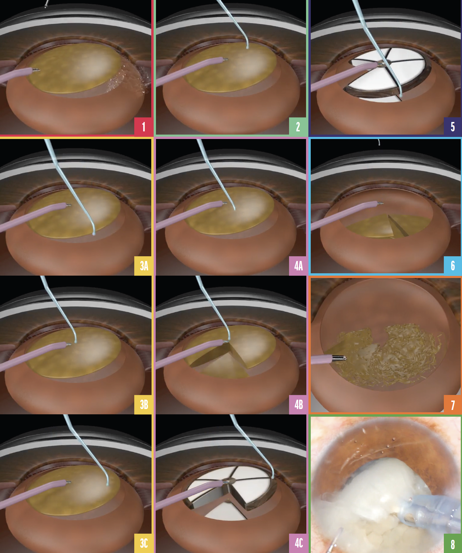
Figure 1. Gentle hydrodissection is used to prolapse the nucleus out of the capsular bag.
Figure 2. A phaco probe is positioned in the center of the nucleus.
Figure 3. A chopper is introduced from the periphery of the nucleus near the 6 clock position (A), brought toward the phaco probe (B), and then rotated toward the 3 clock position (C).
Figure 4. With the phaco chopper at the 3 clock position, the nucleus is tilted and rotated clockwise until the chopper reaches the 6 clock position (A), freeing one-fifth of the nucleus (B and C).
Figure 5. A straight line to the center of the nucleus is drawn with the phaco chopper, freeing the second fifth of the nucleus.
Figure 6. The same steps are repeated until the entire nucleus has been chopped in the supracapsular space.
Figure 7. The nuclear pieces are pushed back into the capsular bag with an OVD and emulsified.
Figure 8. The cartwheel chop technique is performed on a rock-hard cataract.
One alternative with the cartwheel technique is to complete either a partial trench (ie, the first half of a stop and chop technique) or a crater and then impale the probe in the center of the half-trench or crater. The cartwheel technique is then performed.
CONCLUSION
Cartwheel chop is a bridge between stop and chop and direct chop procedures. Executing the chop at the iris plane and phacoemulsification in the bag (Figure 8) can prevent both posterior and endothelial complications.
