


A 36-year-old man presented with pain, redness, and reduced vision in the right eye for 3 days following a mud injury to the eye while working in fields. Anterior segment examination revealed conjunctival congestion, a central, dry-looking 4 x 4 mm ring-shaped anterior to midstromal infiltrate with feathery margins and surrounding satellite lesions (Figure). Fundoscopy was deferred due to the patient’s pain. Visual acuity in the affected eye was 5/60. Corneal scraping was performed for smear examination on a 10% potassium hydroxide wet mount for Gram staining and blood agar, chocolate agar, and sabouraud dextrose agar culture. The smear examination revealed long, slender hyaline hyphae with sparse septations. A diagnosis of fungal keratitis was made.
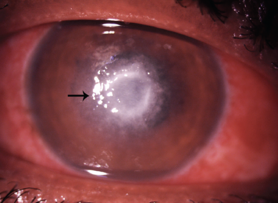
Figure. A central ring-shaped anterior to midstromal infiltrate with feathery margins and surrounding satellite lesions (arrow).
The patient was started on topical antifungal therapy with hourly natamycin 1% and voriconazole 1% and adjuvant therapy. Three days later—to our surprise—the ulcer had worsened, and visual acuity had deteriorated to 2/60.
A diagnosis of Pythium insidiosum keratitis was therefore considered and confirmed with blood agar culture and the leaf incarnation method.1 The patient was switched to topical antibacterial therapy with hourly linezolid 0.2% and azithromycin 1%.2 After 2 weeks of therapy, rapid improvement with signs of healing was evident, and visual acuity improved to 6/60. After 4 weeks, visual acuity had improved to 6/9, and the antibacterial drug regimen was tapered.
This is an interesting and rare case of vision-threatening Pythium insidiosum keratitis masquerading as fungal keratitis. The important clinical finding was the presence of small tentacular projections that masquerade as satellite lesions. A high index of clinical suspicion and expertise is needed to diagnose these cases before culture results are available. This helps prevent the rapid progression of ulcers leading to descemetocele, perforation, and blindness and minimizes the need for keratoplasty.3
1. Gurnani B, Kaur K, Venugopal A, et al. Pythium insidiosum keratitis – A review. Indian J Ophthalmol. 2022;70(4):1107-1120.
2. Maeno S, Oie Y, Sunada A, et al. Successful medical management of Pythium insidiosum keratitis using a combination of minocycline, linezolid, and chloramphenicol. Am J Ophthalmol Case Rep. 2019;15:100498.
3. Gurnani B, Narayana S, Christy J, Rajkumar P, Kaur K, Gubert J. Successful management of pediatric pythium insidiosum keratitis with cyanoacrylate glue, linezolid, and azithromycin: rare case report. Eur J Ophthalmol. 2022;32(5):NP87-NP91.


