


Novel therapeutic keratopigmentation techniques that aim to restore or change the appearance of the disabled and cosmetically abnormal appearance of the cornea in pathologic eyes are emerging. Currently, the available solutions for these eyes, including contact lenses, ocular and orbital prosthetic devices, and penetrating keratoplasty, all have considerable downsides.1 Contact lenses are usually tolerated poorly by patients with corneal and ocular surface diseases, and prostheses are not always well accepted. Waiting lists are long for penetrating keratoplasty. Additionally, it is unethical to perform the procedure for purely cosmetic reasons with patients waiting for corneal transplant to restore vision. The sad truth is that eyes with corneal cosmetic disability often have an uncertain future and limited therapeutic possibilities.
Looking ahead, it may become the standard of care for pathologic eyes to undergo either intrastromal/femtosecond laser–assisted or superficial/manual therapeutic keratopigmentation. I have performed both as standalone procedures and in combination with either oculoplastic or strabismus surgery for more than 14 years with excellent results (see two case presentations later in this article).
The instrumentation for the procedure typically includes the following:
- Micronized mineral pigments;
- A stromal dissector;
- A pupil marker;
- A superficial automated keratopigmentation device (Biotic Phocea); and
- A femtosecond laser.
CASE PRESENTATIONS
▶ No. 1: Femtosecond laser–assisted intrastromal keratopigmentation with superficial automated keratopigmentation. A 55-year-old woman experienced ocular trauma to her right eye. She lost all light perception with no chance of recovering vision. She had a history of retinal detachment and glaucoma. The patient sought cosmetic restoration of her right eye for years (Figure 1A). An examination of the patient at my clinic found epithelial hyperplasia that was secondary to endothelial cell decompensation. Without intervention, the epithelial hyperplasia would eventually relapse. I decided to perform intrastromal therapeutic keratopigmentation using a mixture of pigments to achieve the right iris color.
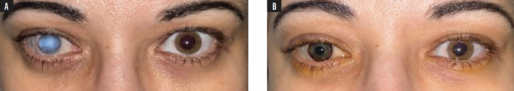
Figure 1. Before (A) and after (B) photographs of the eyes described in case No. 1.
The pupil was made by introducing black pigment first into a pocket created by the femtosecond laser and then superficially. Superficial automated pigmentation was performed to give the iris a greenish appearance that closely matched the iris in the patient’s contralateral eye. Figure 1B shows the eyes at about 1 month after surgery. The patient was happy with her postoperative cosmetic appearance and outcome.
▶ No. 2: Hang-back recession/resection of two horizontal muscles combined with superficial automated keratopigmentation. A 27-year-old man who had experienced an ocular trauma about 20 years earlier presented to my clinic (Figure 2A). He had no light perception and 50.00 PD of exotropia. Treatment for the exotropia and keratopigmentation were initiated simultaneously.
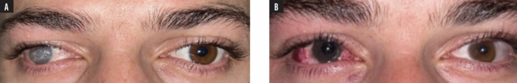
Figure 2. Before (A) and after (B) photographs of the eyes described in case No.2.
The squint surgery was performed first to avoid permanently staining the Tenon capsule. After lateral and middle rectus hang-back recession, superficial automated keratopigmentation surgery was performed. The cornea was manually deepithelialized to avoid unnecessary staining of the area with pigment. The pupillary size was calculated using the size of the mesopic pupil of the patient’s contralateral eye. Once the pupil had been re-created, the periphery of the iris was obtained by staining the cornea. A mixture of two different pigments was required to provide an adequate cosmetic appearance. One week after surgery, the colors of the two eyes matched almost perfectly (Figure 2B). The patient was extremely satisfied.
CONCLUSION
Modern therapeutic augmentation provides a systematic, minimally invasive method for corneal restoration. The procedure can be performed safely on eyes that have corneal pathology with almost no adverse reactions and high patient satisfaction.2 Therapeutic augmentation may also be combined with other procedures to provide patients with excellent outcomes.
1. D’Oria F, Abu-Mustafa SK, Alió JL. Cosmetic change of the apparent color of the eye: a review on surgical alternatives, outcomes, and complications. Ophthalmol Ther. 2022;11(2):465-477.
2. Alio JL, Al-Shymali O, Amesty MA, Rodriguez AE. Keratopigmentation with micronised mineral pigments: complications and outcomes in a series of 234 eyes. Br J Ophthalmol. 2018;102:742-747.
